. 2017 Sep 1;429(1):356-369.
doi: 10.1016/j.ydbio.2017.04.011. Epub 2017 Apr 25.

Migration pathways of sacral neural crest during development of lower urogenital tract innervation

Affiliations

Migration pathways of sacral neural crest during development of lower urogenital tract innervation

Carrie B Wiese et al. Dev Biol. .

Abstract

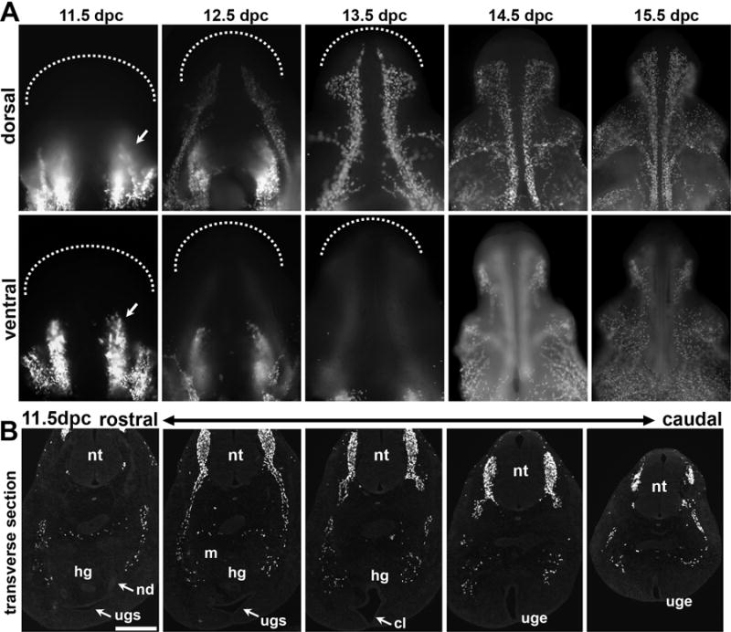
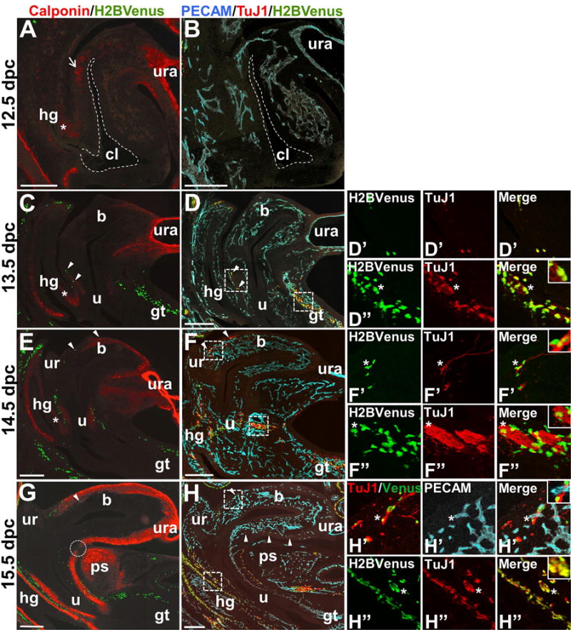
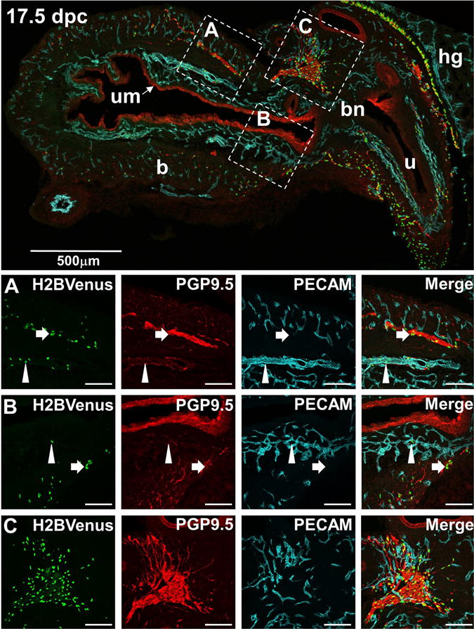
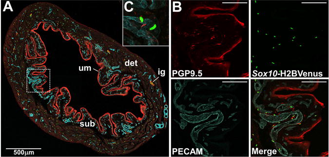
The migration and fate of cranial and vagal neural crest-derived progenitor cells (NCPCs) have been extensively studied; however, much less is known about sacral NCPCs particularly in regard to their distribution in the urogenital system. To construct a spatiotemporal map of NCPC migration pathways into the developing lower urinary tract, we utilized the Sox10-H2BVenus transgene to visualize NCPCs expressing Sox10. Our aim was to define the relationship of Sox10-expressing NCPCs relative to bladder innervation, smooth muscle differentiation, and vascularization through fetal development into adulthood. Sacral NCPC migration is a highly regimented, specifically timed process, with several potential regulatory mileposts. Neuronal differentiation occurs concomitantly with sacral NCPC migration, and neuronal cell bodies are present even before the pelvic ganglia coalesce. Sacral NCPCs reside within the pelvic ganglia anlagen through 13.5 days post coitum (dpc), after which they begin streaming into the bladder body in progressive waves. Smooth muscle differentiation and vascularization of the bladder initiate prior to innervation and appear to be independent processes. In adult bladder, the majority of Sox10+ cells express the glial marker S100β, consistent with Sox10 being a glial marker in other tissues. However, rare Sox10+ NCPCs are seen in close proximity to blood vessels and not all are S100β+, suggesting either glial heterogeneity or a potential nonglial role for Sox10+ cells along vasculature. Taken together, the developmental atlas of Sox10+ NCPC migration and distribution profile of these cells in adult bladder provided here will serve as a roadmap for future investigation in mouse models of lower urinary tract dysfunction.

Keywords: Autonomic nervous system; Bladder; Lower urinary tract; Pelvic ganglia; Peripheral nervous system; Sacral neural crest; Sox10.


Figures

Figure 1
Figure 1. Distribution of sacral neural crest-derived progenitor cells (NCPCs) in and Sox10-H2BVenus embryos
Ventral views of micro-dissected urogenital tracts are shown in whole mount from (A) a 14.5 dpc Sox10LacZ-KO/+ and (B) a 14.5 dpc Sox10-H2BVenus embryo (70× magnification). The superior (anterior) surface of the genital tubercle is shown. Abbreviations: a, adrenal gland; b, bladder; cg, celiac ganglia; dn, dorsal nerve; e, epididymis; gt, genital tubercle; hg, hindgut; k, kidney; t, testis; vd, vas deferens.
Figure 2
Figure 2. Initial migration of sacral NCPCs towards the the developing urogenital tract viewed in whole mount Sox10-H2BVenus embryos
(A) Lateral view of a 10 dpc embryo imaged for H2BVenus fluorescence. The dorsal surface of the tail is outlined with a dashed line, and the most caudal neural crest cells are visible as punctate spots that are just emerging from the neural tube at the level of the hindlimb and are marked with an arrow. (B) Fluorescence image of H2BVenus-expressing NCPC entering the genital tubercle and pelvic mesenchyme of a 11 dpc embryo at the lumbosacral level (53× magnification). (C) Sox10-H2BVenus+ progenitors are visible in this lateral view of a micro-dissected lower urinary tract. The bladder is located above the genital tubercle and out of view behind the umbilical blood vessel in a 12 dpc embryo (50× magnification). (D) Lateral view of the urogenital system micro-dissected from a 12.5 dpc embryo shows Sox10+ NCPC migrating through the the pelvic plexus, prominent aggregation of cells within the pelvic ganglia anlagen (red circle), and migration of sacral NCPCs into the genital tubercle. The dashed line indicates the distal aspect of the genital tubercle. The dotted lines in (B), (C) and (D) denote the paths of umbilical blood vessels that flank the bladder. Abbreviations: e, eye; tg, trigeminal ganglion; o, otic vesicle; drg, dorsal root ganglia; gt, genital tubercle; pg, pelvic ganglion; pp, pelvic plexus.
Figure 3
Figure 3. Sacral NCPCs route around the metanephric mesenchyme and form loose aggregates in the pelvic ganglia anlagen by 12 dpc
(A) Schematic diagram through the mid-sagital fetal LUT at 11.25 dpc illustrates the location of structures in the developing urogenital sinus. The metanephric mesenchyme is lateral to the midline (pink). A red line indicates the level of the plane of sections collected through the developing embryo that are shown in panel B. (B) Transverse cryosection through an 11.25 dpc Sox10-H2BVenus embryo at the level of the hindlimb stained with DAPI shows the anatomy of the metanephric mesenchyme (dashed circle) and the junction of the cloacal cavity with the hindgut. (B′) Immunostaining of the section in panel A with antibodies for HuC/D (neuronal cell bodies, blue) and TuJ1 (neuronal processes, red) shows the relative position of migrating Sox10-H2BVenus+ NCPC (green) (100× magnification). (B″) High magnification image (200×) of boxed area in A″ shows HuC/D+ cells emphasized by arrows. (C) Schematic diagram through the mid-sagital fetal LUT at 12 dpc illustrates the position of the forming primitive bladder and the rise of the metanephric mesenchyme (pink) relative to the hindgut. A red line indicates the plane of section shown in panel D. (D). Transverse cryosection through the sacral region of a 12 dpc Sox10-H2BVenus embryo at an axial level comparable to that shown in panel A. DAPI stain at this stage shows extension of nerve tracts from the neural tube and septation of the hindgut and cloaca. (D′) Immunostaining of the section in panel B with anti-HuC/D and TuJ1 antibodies reveals aggregates of NCPC co-mingled with HuC/D+ cells that are differentiating neurons (100× magnification). (D″) Magnification (200×) of boxed area in panel B′. Abbreviations: A, anterior; CL, cloaca; D, dorsal; drg, dorsal root ganglia; GT, genital tubercle; gtm, genital tubercle mesenchyme; hg, hindgut; m, metanephric mesenchyme; n, nerve tract; nt neural tube; P, posterior; PBL, primitive bladder; pc, peritoneal cavity; ugm, urogenital membrane; UGS, urogenital sinus; upe, urethral plate epithelium; ur, ureter; urs, urorectal septum, V, ventral. Scale bars = 100 microns. Schematic diagrams in Panels A and C were modified from Georgas et al., 2015.
Figure 4
Figure 4. Sacral NCPC populate the primitive bladder by migration from the pelvic ganglia out into the bladder body
Fetal bladder tissue micro-dissected from 12.5 dpc to 15.5 dpc Sox10-H2BVenus embryos was laid flat and imaged by fluorescent stereomicroscopy from the anterior aspect (4×–10× magnification). NCPC are labeled by bright fluorescence of the Sox10-H2BVenus transgene in the pelvic ganglia anlagen and visible as individual discrete cells that migrate out into the bladder body by 14.5 dpc due to nuclear localization of the H2BVenus reporter. Dashed lines delineate the blood vessels flanking the urogenital sinus/bladder at 12.5 and 13.5 dpc. The bladder dome is apparent in the 14.5 dpc and 15.5 dpc bladders.
Figure 5
Figure 5. Stereotypical patterns of Sox10+ sacral NCPC migration throughout the developing genital tubercle
(A) Wholemount Images of the dorsal (superior) surfaces of micro-dissected genital tubercle from Sox10-H2BVenus embryos at each developmental time point from 11.5 through 15.5 dpc are shown across the top row with ventral (inferior) surfaces shown immediately below. Arrows in panels for 11.5 dpc wholemount images indicate the position of ventral columnar aggregates that are pronounced at 11.5dpc and dissipate by 13.5 dpc. Dashed lines delineate the distal tip of the genital tubercle. (100×–150× magnification) (B) Fluorescent images of transverse sections through the caudal aspect of the sacral embryo show the relative locations of Sox10-H2BVenus+ NCPC that migrate around the metanephric mesenchyme (m) towards the urogenital sinus (ugs) in more rostral sections (right). Dispersed groups of Sox10+ cells at 11.5 dpc have not yet reached the flanks of the forming cloaca where the pelvic ganglia will form (middle). In more caudal sections (left) streams of H2BVenus+ cells are visible within the genital tubercle as it extends from the body wall marked by the urogenital epithelium (uge) at the midline. Individual sections are 18 microns with the entire span from the most rostral section to the most caudal section totaling approximately 550 microns. Scale bar = 200 microns.
Figure 6
Figure 6. Neuronal-glial lineage divergence is ongoing within the pelvic ganglia at 14.5 dpc
The relative size and position of a fetal pelvic ganglia in relation to the urinary bladder is shown in a cryosection (top, 100× magnification), where Sox10+ NCPC are labeled by intense H2BVenus expression. Below confocal images of cryosections immunostained with antibodies against BFAPF (red, early glial marker, left column panels) identify Sox10-H2BVenus+ cells (green, A) that exhibit co-localization in merged images (arrows, A″). There are some Sox10+ nuclei that do not show any BFABP co-localization (arrowheads, A″). At 14.5 dpc fewer Sox10-H2BVenus+ show co-localization with the more mature marker of peripheral glial S100b (red, middle column panels, B–B″) in cryosections stained for this antigen (arrows, B′), while most H2BVenus+ cells show no S100b labeling (arrowheads, B″). In contrast, confocal images of pelvic ganglia stained for PGP9.5 (red, neuronal marker, right column panels C – C″) reveal limited co-localization with this marker due to residual perdurance of the Sox10-H2BVenus+ transgene in cells that have progressed towards the neuronal lineage (arrows, C′), while there are many Sox10-H2BVenus+ cells that show no co-localization with PGP9.5 (arrowheads, C″). Confocal magnification is 400× for all images. High magnification insets to illustrate co-localization are show in the upper right of panels A″, B″, and C″. Abbreviations: b, bladder; pg, pelvic ganglia, um, urothelium; ura, urachus. Scale bars in panels A–C″ = 50 microns.
Figure 7
Figure 7. Temporal and spatial independence of sacral NCPC migration marked by Sox10-H2BVenus expression relative to smooth muscle and vasculature development in the bladder wall
Images of sagittal cryosections through the developing lower urogenital tract of 12.5 dpc through 15.5 dpc Sox10-H2BVenus+ embryos are shown. Sox10-expressing NCPCs are visible as a result of H2BVenus transgene fluorescence (green) in sections immunostained either for calponin (red, panels A, C, E, G) to detect smooth muscle alone or jointly immunostained for PECAM (blue, endothelial cells) and TuJ1 (red, neuronal processes) in panels B, D, F, H. In all images, the ventral aspect of the embryo is on the right. Cryosections immunostained for PECAM and TuJ1 were approximately 160um lateral to the midsagittal sections used to detect calponin. For this reason, some midline structures are not visible in the sections used for PECAM/TuJ1 detection. All fluorescent (calponin) and confocal (PECAM/TuJ1) images were taken at 100× magnification and tiled in Adobe Photoshop to generate the composite images shown. An asterisk indicates the position of the extending urorectal septum that separates the hindgut from the cloaca (dashed outline, panels A, B) where smooth muscle differentiation marked by calponin immunoreactivity appears initially. A dotted circle emphasizes a gap in the continuity of calponin staining at the junction between the bladder neck and urethra at 15.5 dpc. Arrowheads indicate the locations of individual Sox10+ NCPCs that are integral to the urethral and bladder walls. High magnification insets in the rightmost column coincide with boxed insets in panels D, F, and H. At 13.5 dpc Abbreviations: b, bladder; cl, cloaca (dashed outline); gt, genital tubercle; hg, hindgut; ps, developing pubic symphysis; u, urethra; ur, ureter; ura, urachus. Scale bars = 200 microns.
Figure 8
Figure 8. Association of NCPCs with both neuronal processes and blood vessels in the 17.5 dpc developing bladder
The top panel shows a low-magnification (70×) midsagittal view of an E17.5 bladder from an Sox10-H2BVenus (green) fetal mouse immunostained with the neuronal marker PGP9.5 (red) and the endothelial marker PECAM (blue). Images in Rows A, B, and C are higher magnification views (200×) of the corresponding areas delineated by dashed squares in the top panel. Typical Sox10-H2BVenus+ glial cells intimately associated with neuronal processes are highlighted by arrows in Rows A and B. Rare Sox10+ cells closely associated with vascular endothelium and distant from neuronal processes are highlighted by arrowheads in Row A and B. A high density of Sox10+ cells, neuronal processes, and vascular endothelium is present within the pelvic ganglion shown in Row C. This tight aggregate of cells precludes an assessment of whether a subpopulation of Sox10+ cells associates more closely with endothelial cells than with neuronal processes. The staining of urothelium with PGP9.5 antibody is consistently observed by many laboratories (See www.gudmap.org Gene Expression Database).
Figure 9
Figure 9. Association of Sox10+ NC-derived cells with vasculature in the adult bladder
(A) Confocal image of a cryosection from adult mouse Sox10-H2BVenus transgenic bladder stained with PECAM (blue, endothelial cells of vasculature) and PGP9.5 (red, neuronal cells and urothelium) shown at 100× magnification. Individual Sox10 expressing cells are labeled by nuclear expression of the H2BVenus reporter (green). (B) Higher zoom confocal images from the boxed region in panel A show individual fluorophore labeling in the bladder wall just beneath the urothelium (200× magnification). (C) High magnification images reveal the presence of Sox10-H2BVenus+ cells whose nuclei are tightly associated with PECAM+ vaculature and appear to wrap around vessels (400× magnification). Abbreviations: det, detrusor muscle; sub, submucosa; um, urothelium. Scale bars = 100 microns.
Figure 10
Figure 10. Heterogeneity of Sox10+ NC-derived cells in the adult mouse bladder
(A) A low-magnification sagittal view of an adult bladder expressing the Sox10-H2BVenus transgene (4× magnification) shows three anatomic layers, urethra, and bladder dome labeled to provide orientation. The urothelium and submucosa present as convoluted folds in an empty adult bladder to provide room for expansion when filled with urine. An intramural ganglion is shown within the dashed oval. Two additional, smaller intramural ganglia are present along the dorsal aspect of the bladder. Areas of the bladder wall defined by boxed insets are shown on the right at higher magnification (200×) following immunohistochemistry performed with a glial marker (S100β) and a neuronal marker (PGP9.5). (B) In the bladder dome and dorsal wall of the bladder, the majority of Sox10+ cells are S100β+. However, infrequent Sox10+, S100β− cells (arrows) and very rare Sox10−, S100β+ cells (arrowheads) are also present. (C) In the detrusor muscle of the bladder dome, there is no colocalization of Sox10-H2BVenus with PGP9.5. However, within the intramural ganglia, there are infrequent Sox10-H2BVenus+ cells that also express PGP9.5 (arrow). A rare Sox10+, PGP9.5+ cell is present near the ganglion (arrowhead). (D) Sox10+ cells are in close proximity but do not colocalize with HuC/D+ large neuronal nuclei that are present in small groups of two to five cells within intramural ganglia (left) or present as individual cells in the bladder wall (right). Abbreviations: ser, serosa; det, detrusor muscle; um, urothelium; ig, intramural ganglion. Scale bars Panels B and C = 100 microns. Scale bars panel D = 20 microns

Similar articles

Cited by

References

    1. Anderson RB, Stewart AL, Young HM. Phenotypes of neural-crest-derived cells in vagal and sacral pathways. Cell Tissue Res. 2006;323:11–25. - PubMed
    1. Atala A. Tissue engineering of human bladder. Br Med Bull. 2011;97:81–104. - PubMed
    1. Atala A, Bauer SB, Soker S, Yoo JJ, Retik AB. Tissue-engineered autologous bladders for patients needing cystoplasty. Lancet. 2006;367:1241–1246. - PubMed
    1. Betancur P, Bronner-Fraser M, Sauka-Spengler T. Genomic code for Sox10 activation reveals a key regulatory enhancer for cranial neural crest. Proc Natl Acad Sci U S A. 2010;107:3570–3575. - PMC - PubMed
    1. Betancur P, Sauka-Spengler T, Bronner M. A Sox10 enhancer element common to the otic placode and neural crest is activated by tissue-specific paralogs. Development. 2011;138:3689–3698. - PMC - PubMed

Publication types

MeSH terms

Substances

LinkOut - more resources