

Zuckerkandl Tubercle of the Thyroid Gland: Correlations between Findings of Anatomic Dissections and CT Imaging
- PMID: 28450435
- PMCID: PMC7959921
- DOI: 10.3174/ajnr.A5172
Zuckerkandl Tubercle of the Thyroid Gland: Correlations between Findings of Anatomic Dissections and CT Imaging
Abstract
Background and purpose: The Zuckerkandl tubercle is located at the posteromedial border of the thyroid lobe, and it may be confused with a neoplasm or other mass. This study was performed to clarify the position and morphologic characteristics of the Zuckerkandl tubercle by dissecting cadavers and to compare the findings with the corresponding CT images obtained in the same cadavers.
Materials and methods: One hundred thyroid lobes from 50 fresh cadavers were dissected for this study (20 males and 30 females; mean age at death, 77.3 ± 11.5 years). CT scans were obtained in 10 of the cadavers by using a 128-channel multidetector row CT scanner before dissection.
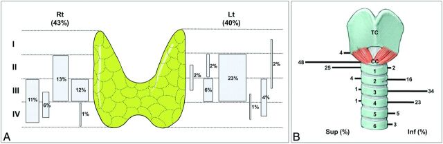
Results: The Zuckerkandl tubercle of the thyroid gland was observed in 83% of the specimens. It was mostly located at the posteromedial border of the thyroid lobe and within the middle two quarters (2nd and 3rd) of the thyroid lobe. The Zuckerkandl tubercle was classified into 3 types based on its direction of extension: posteromedial in 64% of the specimens, posteromedial and superior in 13%, and posteromedial and inferior in 6%. On axial CT, the Zuckerkandl tubercle was usually continuous with the posteromedial part of the thyroid lobe and extended posteromedially to the esophagus. The parts of the Zuckerkandl tubercle that protrude posteromedially and superiorly or posteromedially and inferiorly from the thyroid lobe appeared separated from the thyroid gland by a thin, low-density string on axial CT.
Conclusions: Zuckerkandl tubercles that protrude toward the posteromedial and superior or inferior direction could cause confusion due to their separation when performing diagnoses with CT images.
© 2017 by American Journal of Neuroradiology.
Figures


Similar articles
-
Zuckerkandl's tubercle of the thyroid gland: Its location in the anatomical position, and comparative morphology of the same specimens before and after fixation.Clin Anat. 2015 May;28(4):472-6. doi: 10.1002/ca.22533. Epub 2015 Apr 2. Clin Anat. 2015. PMID: 25832846
-
[Zuckerkandl tubercle of the thyroid gland (anatomo-surgical study: preliminary considerations)].Chir Ital. 1989 Apr-Jun;41(2-3):129-36. Chir Ital. 1989. PMID: 2638217 Italian.
-
The Zuckerkandl tubercle: problematic or helpful in thyroid surgery?Eur Arch Otorhinolaryngol. 2013 Aug;270(8):2327-32. doi: 10.1007/s00405-012-2334-7. Epub 2013 Jan 12. Eur Arch Otorhinolaryngol. 2013. PMID: 23315185
-
Evaluation of thyroid Zuckerkandl tubercle by computed tomography.Surg Radiol Anat. 2022 Jun;44(6):907-912. doi: 10.1007/s00276-022-02963-2. Epub 2022 Jun 6. Surg Radiol Anat. 2022. PMID: 35666298 Review.
-
Zuckerkandl's tubercle and its relationship to the recurrent laryngeal nerve: A cadaveric dissection and meta-analysis.Auris Nasus Larynx. 2017 Dec;44(6):639-647. doi: 10.1016/j.anl.2017.03.013. Epub 2017 Apr 2. Auris Nasus Larynx. 2017. PMID: 28377109 Review.
Cited by
-
Introducing a Pole Concept for Nodule Growth in the Thyroid Gland: Taller-than-Wide Shape, Frequency, Location and Risk of Malignancy of Thyroid Nodules in an Area with Iodine Deficiency.J Clin Med. 2022 May 1;11(9):2549. doi: 10.3390/jcm11092549. J Clin Med. 2022. PMID: 35566675 Free PMC article.
-
Factors Affecting Thyroid Volume in Children Aged 4 to 18 Years.Diagnostics (Basel). 2025 Aug 7;15(15):1980. doi: 10.3390/diagnostics15151980. Diagnostics (Basel). 2025. PMID: 40804944 Free PMC article.
-
A Rare Variant of Zuckerkandl Tubercle: Thyroid Ring.Indian J Otolaryngol Head Neck Surg. 2023 Dec;75(4):4090-4092. doi: 10.1007/s12070-023-04079-4. Epub 2023 Jul 15. Indian J Otolaryngol Head Neck Surg. 2023. PMID: 37974716 Free PMC article.
-
Pediatric hyperparathyroidism: review and imaging update.Pediatr Radiol. 2021 Jun;51(7):1106-1120. doi: 10.1007/s00247-021-05050-7. Epub 2021 Apr 27. Pediatr Radiol. 2021. PMID: 33904951 Review.
-
The clinical significance of remnant thyroid tissue in thyroidectomized differentiated thyroid cancer patients on 131I-SPECT/CT.BMC Med Imaging. 2021 May 8;21(1):78. doi: 10.1186/s12880-021-00612-5. BMC Med Imaging. 2021. PMID: 33964885 Free PMC article.
References
-
- Zuckerkandl E. Nebst Bemerkungen über die Epithelkörperchen des Menschen. Anat Hefte 1902;LXI:61–82
-
- Gilmour JR. The gross anatomy of the parathyroid glands. J Pathol Bacteriol 1938;46:133–49
MeSH terms
LinkOut - more resources
Full Text Sources
Other Literature Sources