Noninvasive Assessment of IDH Mutational Status in World Health Organization Grade II and III Astrocytomas Using DWI and DSC-PWI Combined with Conventional MR Imaging
- PMID: 28450436
- PMCID: PMC7960080
- DOI: 10.3174/ajnr.A5171
Noninvasive Assessment of IDH Mutational Status in World Health Organization Grade II and III Astrocytomas Using DWI and DSC-PWI Combined with Conventional MR Imaging
Abstract
Background and purpose: Isocitrate dehydrogenase (IDH) has been shown to have both diagnostic and prognostic implications in gliomas. The purpose of this study was to examine whether DWI and DSC-PWI combined with conventional MR imaging could noninvasively predict IDH mutational status in World Health Organization grade II and III astrocytomas.
Materials and methods: We retrospectively reviewed DWI, DSC-PWI, and conventional MR imaging in 42 patients with World Health Organization grade II and III astrocytomas. Minimum ADC, relative ADC, and relative maximum CBV values were compared between IDH-mutant and wild-type tumors by using the Mann-Whitney U test. Receiver operating characteristic curve and logistic regression were used to assess their diagnostic performances.
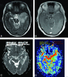
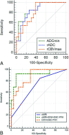
Results: Minimum ADC and relative ADC were significantly higher in IDH-mutated grade II and III astrocytomas than in IDH wild-type tumors (P < .05). Minimum ADC with the cutoff value of ≥1.01 × 10⁻³ mm2/s could differentiate the mutational status with a sensitivity, specificity, positive predictive value, and negative predictive value of 76.9%, 82.6%, 91.2%, and 60.5%, respectively. The threshold value of <2.35 for relative maximum CBV in the prediction of IDH mutation provided a sensitivity, specificity, positive predictive value, and negative predictive value of 100.0%, 60.9%, 85.6%, and 100.0%, respectively. A combination of DWI, DSC-PWI, and conventional MR imaging for the identification of IDH mutations resulted in a sensitivity, specificity, positive predictive value, and negative predictive value of 92.3%, 91.3%, 96.1%, and 83.6%.
Conclusions: A combination of conventional MR imaging, DWI, and DSC-PWI techniques produces a high sensitivity, specificity, positive predictive value, and negative predictive value for predicting IDH mutations in grade II and III astrocytomas. The strategy of using advanced, semiquantitative MR imaging techniques may provide an important, noninvasive, surrogate marker that should be studied further in larger, prospective trials.
© 2017 by American Journal of Neuroradiology.
Figures


Similar articles
-
Predicting 1p/19q codeletion status using diffusion-, susceptibility-, perfusion-weighted, and conventional MRI in IDH-mutant lower-grade gliomas.Acta Radiol. 2021 Dec;62(12):1657-1665. doi: 10.1177/0284185120973624. Epub 2020 Nov 22. Acta Radiol. 2021. PMID: 33222488
-
Prediction of Ki-67 labeling index, ATRX mutation, and MGMT promoter methylation status in IDH-mutant astrocytoma by morphological MRI, SWI, DWI, and DSC-PWI.Eur Radiol. 2023 Oct;33(10):7003-7014. doi: 10.1007/s00330-023-09695-w. Epub 2023 May 3. Eur Radiol. 2023. PMID: 37133522
-
Static 18F-FET PET and DSC-PWI based on hybrid PET/MR for the prediction of gliomas defined by IDH and 1p/19q status.Eur Radiol. 2021 Jun;31(6):4087-4096. doi: 10.1007/s00330-020-07470-9. Epub 2020 Nov 19. Eur Radiol. 2021. PMID: 33211141
-
The T2-FLAIR-mismatch sign as an imaging biomarker for IDH and 1p/19q status in diffuse low-grade gliomas: a systematic review with a Bayesian approach to evaluation of diagnostic test performance.Neurosurg Focus. 2019 Dec 1;47(6):E13. doi: 10.3171/2019.9.FOCUS19660. Neurosurg Focus. 2019. PMID: 31786548
-
Diagnostic accuracy and potential covariates for machine learning to identify IDH mutations in glioma patients: evidence from a meta-analysis.Eur Radiol. 2020 Aug;30(8):4664-4674. doi: 10.1007/s00330-020-06717-9. Epub 2020 Mar 19. Eur Radiol. 2020. PMID: 32193643 Review.
Cited by
-
Noninvasive Determination of the IDH Status of Gliomas Using MRI and MRI-Based Radiomics: Impact on Diagnosis and Prognosis.Curr Oncol. 2022 Sep 23;29(10):6893-6907. doi: 10.3390/curroncol29100542. Curr Oncol. 2022. PMID: 36290819 Free PMC article. Review.
-
Diffusion Weighted Imaging in Neuro-Oncology: Diagnosis, Post-Treatment Changes, and Advanced Sequences-An Updated Review.Cancers (Basel). 2023 Jan 19;15(3):618. doi: 10.3390/cancers15030618. Cancers (Basel). 2023. PMID: 36765575 Free PMC article. Review.
-
A radiomics nomogram may improve the prediction of IDH genotype for astrocytoma before surgery.Eur Radiol. 2019 Jul;29(7):3325-3337. doi: 10.1007/s00330-019-06056-4. Epub 2019 Apr 10. Eur Radiol. 2019. PMID: 30972543
-
Magnetic Resonance Imaging Derived Biomarkers of IDH Mutation Status and Overall Survival in Grade III Astrocytomas.Diagnostics (Basel). 2020 Apr 23;10(4):247. doi: 10.3390/diagnostics10040247. Diagnostics (Basel). 2020. PMID: 32340318 Free PMC article.
-
Comparison of [18F]Fluoroethyltyrosine PET and Sodium MRI in Cerebral Gliomas: a Pilot Study.Mol Imaging Biol. 2020 Feb;22(1):198-207. doi: 10.1007/s11307-019-01349-y. Mol Imaging Biol. 2020. PMID: 30989437
References
-
- Daumas-Duport C, Scheithauer B, O'Fallon J, et al. . Grading of astrocytomas: a simple and reproducible method. Cancer 1988;62:2152–65 - PubMed
MeSH terms
Substances
LinkOut - more resources
Full Text Sources
Other Literature Sources
Medical
