Multicenter Study
. 2017 Jun;38(6):1103-1110.
doi: 10.3174/ajnr.A5227. Epub 2017 Apr 27.

Synthetic MRI for Clinical Neuroimaging: Results of the Magnetic Resonance Image Compilation (MAGiC) Prospective, Multicenter, Multireader Trial

Affiliations
Multicenter Study

Synthetic MRI for Clinical Neuroimaging: Results of the Magnetic Resonance Image Compilation (MAGiC) Prospective, Multicenter, Multireader Trial

L N Tanenbaum et al. AJNR Am J Neuroradiol. 2017 Jun.

Abstract

Background and purpose: Synthetic MR imaging enables reconstruction of various image contrasts from 1 scan, reducing scan times and potentially providing novel information. This study is the first large, prospective comparison of synthetic-versus-conventional MR imaging for routine neuroimaging.

Materials and methods: A prospective multireader, multicase noninferiority trial of 1526 images read by 7 blinded neuroradiologists was performed with prospectively acquired synthetic and conventional brain MR imaging case-control pairs from 109 subjects (mean, 53.0 ± 18.5 years of age; range, 19-89 years of age) with neuroimaging indications. Each case included conventional T1- and T2-weighted, T1 and T2 FLAIR, and STIR and/or proton density and synthetic reconstructions from multiple-dynamic multiple-echo imaging. Images were randomized and independently assessed for diagnostic quality, morphologic legibility, radiologic findings indicative of diagnosis, and artifacts.

Results: Clinical MR imaging studies revealed 46 healthy and 63 pathologic cases. Overall diagnostic quality of synthetic MR images was noninferior to conventional imaging on a 5-level Likert scale (P < .001; mean synthetic-conventional, -0.335 ± 0.352; Δ = 0.5; lower limit of the 95% CI, -0.402). Legibility of synthetic and conventional morphology agreed in >95%, except in the posterior limb of the internal capsule for T1, T1 FLAIR, and proton-density views (all, >80%). Synthetic T2 FLAIR had more pronounced artifacts, including +24.1% of cases with flow artifacts and +17.6% cases with white noise artifacts.

Conclusions: Overall synthetic MR imaging quality was similar to that of conventional proton-density, STIR, and T1- and T2-weighted contrast views across neurologic conditions. While artifacts were more common in synthetic T2 FLAIR, these were readily recognizable and did not mimic pathology but could necessitate additional conventional T2 FLAIR to confirm the diagnosis.


Figures

Fig 1.
Fig 1.
Axial synthetic and conventional 3T MR imaging of a normal brain. Conventional (upper row) and synthetic (lower row) image sets exhibit similar legibility and quality. Slight differences in contrast levels are apparent, which do not adversely impact the diagnostic utility of images, particularly between T1 FLAIR and T2 FLAIR views.
Fig 2.
Fig 2.
Left frontal lobe cystic tumor on axial synthetic and conventional 3T MR imaging in a 31-year-old woman. Conventional (upper row) and synthetic (lower row) image sets exhibit similar legibility and quality.
Fig 3.
Fig 3.
Chronic right cerebellar infarction in a 37-year-old woman on axial synthetic and conventional 3T MR imaging. Conventional (upper row) and synthetic (lower row) image sets exhibit similar legibility and quality.
Fig 4.
Fig 4.
Multiple sclerosis in axial synthetic and conventional 3T MR imaging in a 58-year-old woman. Multifocal demyelinating lesions are apparent within the cerebral white matter; these lesions appear similar on conventional (upper row) and synthetic (lower row) image sets. A slight misregistration is apparent due to patient motion between the MDME scan (used for synthetic reconstruction, lower row) and the comparable conventional scan acquired in the study (upper row). While misregistration due to motion can pose challenges in conventional serial acquisitions due to partial section differences in images across contrast views, synthetic reconstruction inherently prevents misregistration across synthetic contrast views.
Fig 5.
Fig 5.
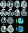
Chronic infarction in synthetic and conventional 3T MR imaging shown alongside color functional perfusion maps in a 62-year-old man. Conventional (rows 1 and 3) and synthetic (rows 2 and 4) views show similar legibility and quality. T1 FLAIR and T2 FLAIR views have some granulated white noise in the margins. Color quantitative perfusion maps (lower right) demonstrate decreased flow and prolonged transit time in this region of chronic infarction.
Fig 6.
Fig 6.
Subdural hematoma on T2 FLAIR in synthetic and conventional 3T MR imaging demonstrating pronounced artifacts. Conventional (left) and synthetic (right) T2 FLAIR images are shown for a patient with subdural hematoma, in which synthetic T2 FLAIR has notable granulated hyperintensities and lacks contrast between the lesion and surrounding tissues. Artifacts of this severity level were rare among synthetically reconstructed images, possibly due to issues in the MDME acquisition that are typically resolved on rescanning. For cases demonstrating these granulated hyperintensities on the synthetic T2 FLAIR, artifacts were readily recognizable by characteristic distortion and correlation with other contrast views without apparent artifacts. While these could necessitate rescanning with conventional T2 FLAIR in some cases, when coupled with other contrast views, these artifacts did not interfere with the diagnostic accuracy of synthetic MR imaging.

References

    1. Riederer S, Suddarth S, Bobman S. Automated MR image synthesis: feasibility studies. Radiology 1984;153:203–06 10.1148/radiology.153.1.6089265 - DOI - PubMed
    1. Warntjes J, Dahlqvist O, Lundberg P. Novel method for rapid, simultaneous T1, T2*, and proton density quantification. Magn Reson Med 2007;57:528–37 10.1002/mrm.21165 - DOI - PubMed
    1. Krauss W, Gunnarsson M, Andersson T, et al. . Accuracy and reproducibility of a quantitative magnetic resonance imaging method for concurrent measurements of tissue relaxation times and proton density. Magn Reson Imaging 2015;33:584–91 10.1016/j.mri.2015.02.013 - DOI - PubMed
    1. Warntjes J, Leinhard O, West J, et al. . Rapid magnetic resonance quantification on the brain: optimization for clinical usage. Magn Reson Med 2008;60:320–29 10.1002/mrm.21635 - DOI - PubMed
    1. Blystad I, Warntjes J, Smedby O, et al. . Synthetic MRI of the brain in a clinical setting. Acta Radiol 2012;53:1158–63 10.1258/ar.2012.120195 - DOI - PubMed

Publication types