. 2017 May 24;37(21):5309-5318.
doi: 10.1523/JNEUROSCI.3472-16.2017. Epub 2017 Apr 27.

Decreased Motor Neuron Support by SMA Astrocytes due to Diminished MCP1 Secretion

Affiliations

Decreased Motor Neuron Support by SMA Astrocytes due to Diminished MCP1 Secretion

Jasmin E Martin et al. J Neurosci. .

Abstract

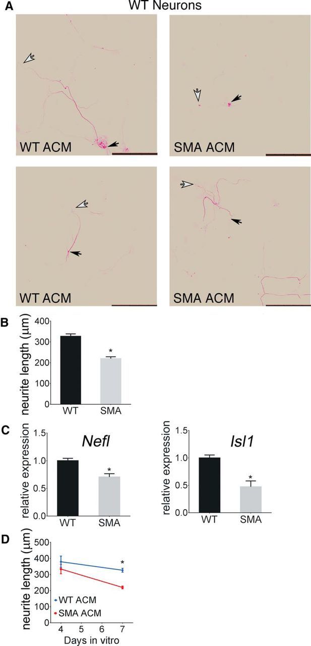
Spinal muscular atrophy (SMA) is an autosomal-recessive disorder characterized by severe, often fatal muscle weakness due to loss of motor neurons. SMA patients have deletions and other mutations of the survival of motor neuron 1 (SMN1) gene, resulting in decreased SMN protein. Astrocytes are the primary support cells of the CNS and are responsible for glutamate clearance, metabolic support, response to injury, and regulation of signal transmission. Astrocytes have been implicated in SMA as in in other neurodegenerative disorders. Astrocyte-specific rescue of SMN protein levels has been shown to mitigate disease manifestations in mice. However, the mechanism by which SMN deficiency in astrocytes may contribute to SMA is unclear and what aspect of astrocyte activity is lacking is unknown. Therefore, it is worthwhile to identify defects in SMN-deficient astrocytes that compromise normal function. We show here that SMA astrocyte cultures derived from mouse spinal cord of both sexes are deficient in supporting both WT and SMN-deficient motor neurons derived from male, female, and mixed-sex sources and that this deficiency may be mitigated with secreted factors. In particular, SMN-deficient astrocytes have decreased levels of monocyte chemoactive protein 1 (MCP1) secretion compared with controls and MCP1 restoration stimulates outgrowth of neurites from cultured motor neurons. Correction of MCP1 deficiency may thus be a new therapeutic approach to SMA.SIGNIFICANCE STATEMENT Spinal muscular atrophy (SMA) is caused by the loss of motor neurons, but astrocyte dysfunction also contributes to the disease in mouse models. Monocyte chemoactive protein 1 (MCP1) has been shown to be neuroprotective and is released by astrocytes. Here, we report that MCP1 levels are decreased in SMA mice and that replacement of deficient MCP1 increases differentiation and neurite length of WT and SMN-deficient motor-neuron-like cells in cell culture. This study reveals a novel aspect of astrocyte dysfunction in SMA and indicates a possible approach for improving motor neuron growth and survival in this disease.

Keywords: MCP1/CCL2; astrocytes; iPSC; motor neuron; spinal muscular atrophy.


Figures

Figure 1.
Figure 1.
Cultured WT mouse spinal cord motor neurons have shorter neurites and express lower levels of motor neuron markers when grown in the presence of SMA ACM compared with WT ACM. Motor neurons were derived from WT mouse E13.5 spinal cords. The neurons were treated 24 h after plating for 7 d with ACM from astrocytes from the spinal cords of either SMA or WT littermate pups. A, Primary neurites identified by SMI-32 immunostaining. Scale bar, 250 μm. The cell soma is indicated by a black arrow and the end of a measured neurite by a white arrow. B, A minimum of 20 neurons were measured and quantified by a blinded evaluator (p = 0.001). C, Relative mRNA levels of Nefl (p = 0.02) and Isl1 (p = 0.002) are reduced in neurons grown in the presence of SMA ACM. D, At 4 d, the neurite lengths are similar between WT and SMA, but at 7 d, the motor neuron neurites in SMA ACM were significantly shorter than the motor neuron neurites grown in WT ACM (p = 0.02). Neurite data are shown as mean ± SEM. Relative mRNA levels are normalized to Pgk1 and mRNA data shown are presented as mean ± SD.
Figure 2.
Figure 2.
Differentiated motor-neuron-like cells from healthy control iPSCs have shorter neurites and express lower levels of motor neuron markers when grown in the presence of SMA ACM compared with WT ACM. iPSCs were differentiated into motor-neuron-like cells. The neurons were treated 24 h after plating for 7 d with ACM from astrocytes from the spinal cords of either SMA or WT littermate pups. A, A minimum of 20 neurons were measured and quantified (p = 0.009). B, Relative mRNA levels of Isl1 (p = 0.001) are reduced in neurons grown in the presence of SMA ACM. Differentiated motor neurons from SMA patient iPSCs have shorter neurites and express lower levels of motor neuron markers when grown in the presence of SMA ACM compared with WT ACM. C, A minimum of 20 neurons were measured and quantified (p = 0.03). D, Relative mRNA levels of TUBB3 (p = 0.02) are reduced in neurons grown in the presence of SMA ACM. Neurite data are shown as mean ± SEM. Relative mRNA levels are normalized to Pgk1 and GusB and mRNA data shown are presented as mean ± SD.
Figure 3.
Figure 3.
MCP1 levels are decreased in SMA ACM compared with WT ACM. Astrocytes were cultured from P2 neonatal mouse spinal cord by enzymatic and mechanical disruption. Once confluent, the cells were incubated with complete astrocyte medium for 5 d. The medium was then collected and tested using a 32 chemokine panel from the University of Maryland Cytokine Core Laboratory. A, Cytokines within detectable limits and with a 20% difference between the WT and SMA average values are shown (protein values in pg/ml). B, secondary validation using a sandwich ELISA for MCP1, RANTES, and IL-6 (blinded). All ELISA data are shown as mean ± SD (p = 0.01).
Figure 4.
Figure 4.
MCP1 mRNA levels are decreased in young SMA mouse spinal cords compared with unaffected littermate controls. MCP1 mRNA levels from spinal cords from P1, P5, and P10 mice were quantified using qPCR by a blinded evaluator. Relative mRNA levels are normalized to Pgk1. Data are shown as mean ± SEM (day 0, p = 0.0004; day 5, p = 0.02).
Figure 5.
Figure 5.
Application of exogenous MCP1 increases the neurite length of cultured WT mouse spinal cord motor neurons. Motor neurons were derived from WT mouse E13.5 spinal cords. The neurons were treated 24 h after plating for 7 d with 500 pg/ml of media recombinant MCP1 or PBS. A, Primary neurites as indicated by SMI-32 immunostaining. A, B, A minimum of 20 neurons were measured and quantified (p = 0.04). Cell soma are indicated by a black arrow and the end of a measured neurite by a white arrow. C, Relative mRNA levels of Isl1 (p = 0.05) are increased in isolated neurons grown in the presence of recombinant MCP1. Neurite data are shown as mean ± SEM. Relative mRNA levels are normalized to Pgk1. mRNA data are shown as mean ± SD.
Figure 6.
Figure 6.
Application of exogenous MCP1 increases the neurite length of motor-neuron-like cells derived from human control and SMA iPSCs. The cells were treated 24 h after plating for 7 d with 500 pg/ml exogenous MCP1 or PBS. A, A minimum of 20 neurons were measured and quantified (p = 0.02). B, Relative mRNA levels of Nefl (p = 0.05) are increased in neurons grown in the presence of recombinant MCP1. Application of exogenous MCP1 increases the neurite length of motor-neuron-like cells derived from SMA patient iPSCs. The cells were treated 24 h after plating for 7 d with 500 pg/ml exogenous MCP1 or PBS. C, A minimum of 20 neurons were measured and quantified (p = 0.002). D, Relative mRNA levels of TUBB3 and Isl1 are unchanged in SMA neurons grown in the presence of recombinant MCP1. Neurite data are shown as mean ± SEM. Relative mRNA levels are normalized to Pgk1 and GusB. mRNA data are shown as mean ± SD.
Figure 7.
Figure 7.
Neutralizing antibody to MCP1 reduced the neurite lengths of cultured WT mouse spinal cord motor neurons. Motor neurons were derived from WT mouse E13.5 spinal cords. The neurons were treated 24 h after plating for 7 d with a neutralizing antibody to MCP1 in WT ACM. A, Primary neurites as indicated by Nefl immunostaining. B, A minimum of 20 neurons were measured and quantified (p = 0.03). Cell soma are indicated by a black arrow and the end of a measured neurite by a white arrow. C, Relative mRNA levels of Nefl (p = 0.0001) and Isl1 (p = 0.0001) are reduced in neurons grown in the presence of WT ACM with the neutralizing MCP1 antibody. D, Relative mRNA levels of Nefl and Isl1 were not different between neurons grown in the presence or absence of a neutralizing antibody to IL-6. Neurite data are shown as mean ± SEM. Relative mRNA levels are normalized to Pgk1. mRNA data are shown as mean ± SD.
Figure 8.
Figure 8.
Neutralizing antibody to MCP1 decreases the neurite length of differentiated motor-neuron-like cells from healthy control and SMA iPSCs. After plating for 7 d, the cells were treated for 24 h with a neutralizing antibody to MCP1 in WT ACM. A, A minimum of 20 neurons were measured and quantified (p = 0.02). B, Nefl relative mRNA level is decreased in the presence of the antibody to MCP1 (p = 0.0001). Neutralizing antibody to MCP1 also decreases the neurite length of differentiated motor-neuron-like cells from SMA patient iPSCs. C, A minimum of 20 neurons were measured and quantified (p = 0.03). D, The TUBB3 relative mRNA level is decreased in the presence of the antibody to MCP1 (p = 0.017). Neurite data are shown as mean ± SEM. Relative mRNA levels are normalized to Pgk1 and GusB. Relative mRNA data are shown as mean ± SD.

Comment in

Similar articles

Cited by

References

    1. Andjelkovic AV, Kerkovich D, Shanley J, Pulliam L, Pachter JS (1999) Expression of binding sites for beta chemokines on human astrocytes. Glia 28:225–235. - PubMed
    1. Araki S, Hayashi M, Tamagawa K, Saito M, Kato S, Komori T, Sakakihara Y, Mizutani T, Oda M (2003) Neuropathological analysis in spinal muscular atrophy type II. Acta Neuropathol 106:441–448. 10.1007/s00401-003-0743-9 - DOI - PubMed
    1. Aït-Ikhlef A, Hantaz-Ambroise D, Henderson CE, Rieger F (2000) Influence of factors secrAieted by wobbler astrocytes on neuronal and motoneuronal survival. J Neurosci Res 59:100–106. - PubMed
    1. Berretta A, Gowing EK, Jasoni CL, Clarkson AN (2016) Sonic hedgehog stimulates neurite outgrowth in a mechanical stretch model of reactive-astrogliosis. Sci Rep 6:21896. 10.1038/srep21896 - DOI - PMC - PubMed
    1. Bianchi LM, Daruwalla Z, Roth TM, Attia NP, Lukacs NW, Richards AL, White IO, Allen SJ, Barald KF (2005) Immortalized mouse inner ear cell lines demonstrate a role for chemokines in promoting the growth of developing statoacoustic ganglion neurons. J Assoc Res Otolaryngol 6:355–367. - PMC - PubMed

Publication types