Review
. 2017 Jun 15;57(6):261-266.
doi: 10.2176/nmc.ra.2017-0043. Epub 2017 Apr 27.

Anomalies of the Middle Cerebral Artery

Affiliations
Review

Anomalies of the Middle Cerebral Artery

Naoyuki Uchiyama. Neurol Med Chir (Tokyo). .

Abstract

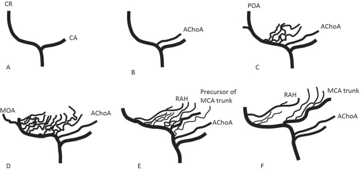
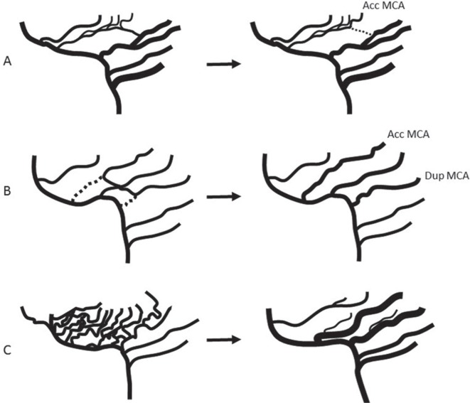
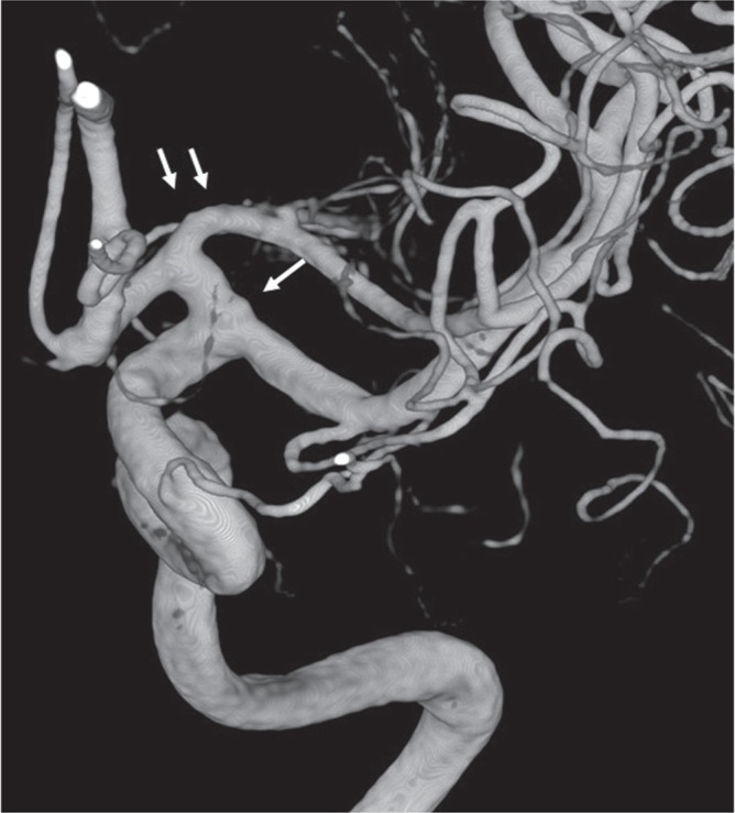
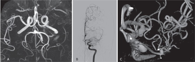
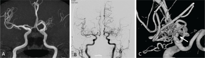
There are several anomalies of the middle cerebral artery (MCA) in humans, such as accessory MCA, duplicated MCA, fenestration of MCA, and duplicated origin of MCA. Recently, unfused or twig-like MCA, which indicates MCA trunk occlusion with collateral plexiform arterial network, have been reported. During the embryonic stage, MCA is thought to generate from plexiform arterial twigs arising from the anterior cerebral artery, and these twigs form the definitive MCA by fusion and regression at the end of the development stage. Any interruption during the fusion of the arterial twigs may result in MCA anomalies, and the unfused or twig-like MCA, especially, is hypothesized to be the persistent primitive arterial twigs. Clinically, it is challenging to differentiate the unfused or twig-like MCA from unilateral moyamoya disease, in which stenotic change begins at the MCA. The knowledge of the anomalies of the MCA is important to perform a safe surgical or endovascular intervention.

Keywords: anatomy; anomaly; embryology; middle cerebral artery.


Conflict of interest statement

Conflicts of Interest Disclosure

The author has no conflicts of interest with regard to this manuscript. The author has registered online Self-reported COI Disclosure Statement Forms.

Figures

Fig. 1
Fig. 1
Schematic representation of the formation of internal cerebral artery (ICA), anterior cerebral artery (ACA), and middle cerebral artery (MCA). A. ICA divides into a cranial branch (CR) and a caudal branch (CA). B. Anterior choroidal artery (AChoA) arises from the CR. C. Multiple plexiform arterial twigs appear just distal to the ACoA. The terminal end of CR constitutes the primitive olfactory artery (POA). D. The POA dwindles. Medial olfactory artery (MOA), which will constitute the ACA, branches out from the POA. E. Multiple plexiform arterial twigs evolve into recurrent artery of Heubner (RAH), medial and lateral striate arteries, and a single MCA trunk. F. Adult configuration of cerebral arteries of anterior circulation.
Fig. 2
Fig. 2
Formation of accessory MCA. A. left: Anastomosis between medial and lateral striate arteries can exist. If communication between MCA trunk and frontal branch of MCA is interrupted (dotted line), accessory (Acc) MCA will be formed. B. If anterior temporal artery arises directly from ICA, it is called duplicated (Dup) MCA. If orbitofrontal artery arises directly from ACA, it is called accessory MCA. C. Multiple plexiform arterial twigs evolve into a single main trunk and lateral striate arteries by fusion and regression. Failure of fusion can form double MCA trunks.
Fig. 3
Fig. 3
Double MCA trunks. If the point of single arrow is considered as the ICA terminal, distal MCA trunk is called accessory MCA. On the other hand, a point of double arrows as ICA terminal, proximal MCA trunk is called duplicated MCA.
Fig. 4
Fig. 4
Case 1. A. MR angiography shows left MCA occlusion. B. Conventional angiography shows twig-like MCA. C. 3D-rotational angiography clearly shows plexiform arterial twigs.
Fig. 5
Fig. 5
Case 2. A. MR angiography shows left MCA occlusion with plexiform arterial twig. B. Conventional angiography shows twig-like MCA. C. 3D-rotational angiography clearly shows plexiform arterial twigs and an aneurysm in the twig (arrow).

References

    1. Lasjaunias P, Berenstein A, ter Brugge KG: Intradural arteries, Surgical Neuroangiography, ed 2 Vol 1 New York, Springer; 2001, pp. 479–630
    1. Crompton MR: The pathology of ruptured middle-cerebral aneurysms with special reference to the differences between the sexes. Lancet 2: 421–425, 1962 - PubMed
    1. Teal JS, Rumbaugh CL, Bergeron RT, Segall HD: Anomalies of the middle cerebral artery: accessory artery, duplication, and early bifurcation. Am J Roentgenol 118: 567–575, 1973 - PubMed
    1. Abanou A, Lasjaunias P, Manelfe C, Lopez-Ibor L: The accessory middle cerebral artery (AMCA). Diagnostic and therapeutic consequences. Anat Clin 6: 305–309, 1984 - PubMed
    1. Takahashi S, Hoshino F, Uemura K, Takahashi A, Sakamoto K: Accessory middle cerebral artery: is it a variant form of the recurrent artery of Heubner? AJNR Am J Neuroradiol 10: 563–568, 1989 - PMC - PubMed

MeSH terms