. 2017 Apr 24:9:42.
doi: 10.1186/s13148-017-0341-7. eCollection 2017.

Aberrant DNA methylation and expression of SPDEF and FOXA2 in airway epithelium of patients with COPD

Affiliations

Aberrant DNA methylation and expression of SPDEF and FOXA2 in airway epithelium of patients with COPD

J Song et al. Clin Epigenetics. .

Abstract

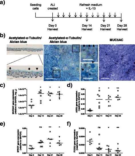
Background: Goblet cell metaplasia, a common feature of chronic obstructive pulmonary disease (COPD), is associated with mucus hypersecretion which contributes to the morbidity and mortality among patients. Transcription factors SAM-pointed domain-containing Ets-like factor (SPDEF) and forkhead box protein A2 (FOXA2) regulate goblet cell differentiation. This study aimed to (1) investigate DNA methylation and expression of SPDEF and FOXA2 during goblet cell differentiation and (2) compare this in airway epithelial cells from patients with COPD and controls during mucociliary differentiation.

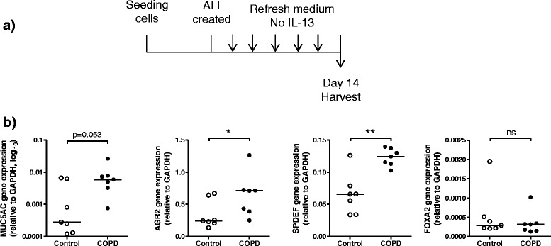
Methods: To assess DNA methylation and expression of SPDEF and FOXA2 during goblet cell differentiation, primary airway epithelial cells, isolated from trachea (non-COPD controls) and bronchial tissue (patients with COPD), were differentiated by culture at the air-liquid interface (ALI) in the presence of cytokine interleukin (IL)-13 to promote goblet cell differentiation.

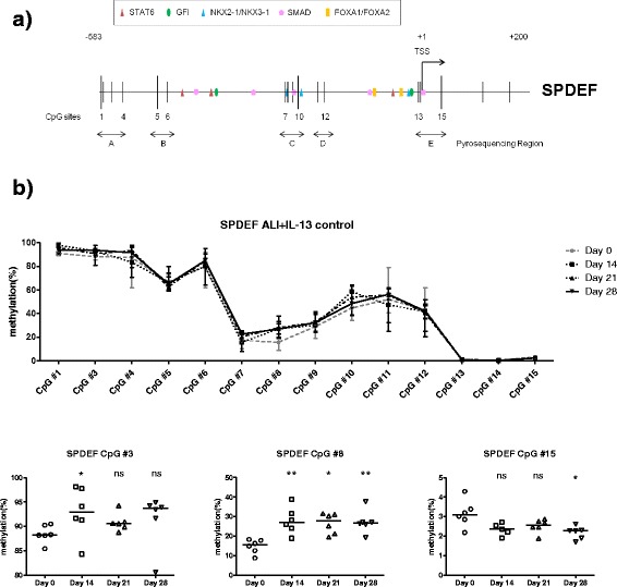
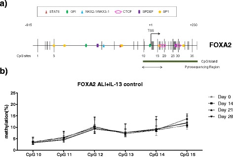
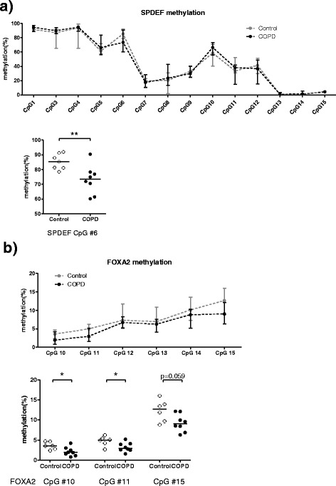
Results: We found that SPDEF expression was induced during goblet cell differentiation, while FOXA2 expression was decreased. Importantly, CpG number 8 in the SPDEF promoter was hypermethylated upon differentiation, whereas DNA methylation of FOXA2 promoter was not changed. In the absence of IL-13, COPD-derived ALI-cultured cells displayed higher SPDEF expression than control-derived ALI cultures, whereas no difference was found for FOXA2 expression. This was accompanied with hypomethylation of CpG number 6 in the SPDEF promoter and also hypomethylation of CpG numbers 10 and 11 in the FOXA2 promoter.

Conclusions: These findings suggest that aberrant DNA methylation of SPDEF and FOXA2 is one of the factors underlying mucus hypersecretion in COPD, opening new avenues for epigenetic-based inhibition of mucus hypersecretion.

Keywords: COPD; DNA methylation; FOXA2; Mucus; SPDEF.


Figures

Fig. 1
Fig. 1
Characterization of the differentiation state of primary bronchial epithelial cells (PBEC) cultured in the air-liquid interface (ALI) model. a Schematic representation of the ALI culture model. PBECs were seeded on to a transwell insert and grown until confluence. Thereafter, apical medium was removed to create an ALI. Cells were harvested after 0, 14, 21, or 28 days for RNA, DNA, and morphology analysis. bd PBEC from control subjects 1–6 (Table 1, n = 6) were cultured at ALI with IL-13 stimulation. b Representative images of immunohistochemistry staining on the differentiated ciliated cells and goblet cells at ALI day 21. Ciliated cells were determined by acetylated-α-tubulin antibody staining and specified by arrows in the images; goblet cells were determined by Alcian Blue staining and MUC5AC antibody staining and were specified by arrow heads in the images. mRNA expressions of c MUC5AC, d AGR2, e SPDEF, and f FOXA2 were analyzed by real-time quantitative PCR at four different time points. Medians are indicated. Significance was tested by the Kruskal-Wallis non-parametric test with Dunn’s posttest data. ns not significant. *p < 0.05; **p < 0.01. Data from days 14, 21, and 28 were compared to day 0
Fig. 2
Fig. 2
Dynamic changes of DNA methylation in the SPDEF promoter during goblet cell differentiation of PBEC from control subjects. a Schematic representation of the promoter region of the SPDEF gene, outlining the putative binding sites for the most relevant transcription factors (STAT6, GFI, NKX2-1/NKX3-1, SMAD, and FOXA1/FOXA2) as analyzed using MatInspector software. The transcription start site was shown as +1. CpGs are indicated as vertical bars. DNA methylation status of 15 CpGs was analyzed using pyrosequencing for the indicated areas. b PBEC from control subjects1–6 (Table 1, n = 6) were cultured at ALI, and DNA methylation levels were analyzed at four different time points. Data represent the connected median methylation levels (min to max) of different CpG sites at each time points, and differential methylated CpG sites are specified with medians indicated. Significance was tested by the Kruskal-Wallis non-parametric test with Dunn’s posttest data. ns not significant. *p < 0.05; **p < 0.01. Data from days 14, 21, and 28 were compared to day 0
Fig. 3
Fig. 3
Dynamic changes of DNA methylation in the FOXA2 promoter during goblet cell differentiation of PBEC from control subjects. a Schematic representations of the promoter region of the FOXA2 gene, outlining the putative binding sites for transcription factors (STAT6, GFI, NKX2-1/NKX3-1, CTCF, SPDEF, and SP1) (MatInspector, top relevants). DNA methylation levels of six CpG sites in the promoter, which were part of a CpG island (indicated in the figure), were examined for the indicated areas. b PBEC from control subjects 1–6 (Table 1, n = 6) were cultured at ALI and DNA methylation levels were analyzed at four different time points. Data represent the connected median methylation levels (min to max) of different CpG sites at each time points
Fig. 4
Fig. 4
Differential mRNA expression profiles in PBECs from COPD patients and control subjects. a Schematic representation of the ALI culture model. PBECs from control 7–17 and COPD 6–16 (Table 1) were seeded onto transwell inserts and grown to confluence. Thereafter, apical medium was removed to create an ALI. Cells were harvested after 14 days for RNA and DNA analysis. b Expressions of MUC5AC, AGR2, SPDEF, and FOXA2 were analyzed by real-time quantitative PCR. Significance was tested by the Mann-Whitney U test. Medians are indicated. ns not significant. *p < 0.05; **p < 0.01
Fig. 5
Fig. 5
Differential DNA methylation of SPDEF and FOXA2 in the PBECs from COPD patients and control subjects. PBECs from control 7–17 and COPD 6–16 (Table 1) were differentiated at ALI for 14 days, and DNA methylation of a SPDEF and b FOXA2 was analyzed by pyrosequencing. Data represent the connected median methylation levels (min to max) of different CpG sites in controls or in COPD, and differential methylated CpG sites between COPD-derived cultures and control-derived cultures are shown. Significance was tested by the Mann-Whitney U test. Medians are indicated. ns not significant. *p < 0.05; **p < 0.01

Similar articles

Cited by

References

    1. Miravitlles M. Cough and sputum production as risk factors for poor outcomes in patients with COPD. Respir Med. 2011;105:1118–28. doi: 10.1016/j.rmed.2011.02.003. - DOI - PubMed
    1. Allinson JP, Hardy R, Donaldson GC, Shaheen SO, Kuh D, Wedzicha JA. The presence of chronic mucus hypersecretion across adult life in relation to chronic obstructive pulmonary disease development. Am J Respir Crit Care Med. 2016;193:662–72. doi: 10.1164/rccm.201511-2210OC. - DOI - PMC - PubMed
    1. Ramos FL, Krahnke JS, Kim V. Clinical issues of mucus accumulation in COPD. Int J Chron Obstruct Pulmon Dis. 2014;9:139–50. - PMC - PubMed
    1. Herriges M, Morrisey EE. Lung development: orchestrating the generation and regeneration of a complex organ. Development. 2014;141:502–13. doi: 10.1242/dev.098186. - DOI - PMC - PubMed
    1. Crystal RG. Airway basal cells. The “smoking gun” of chronic obstructive pulmonary disease. Am J Respir Crit Care Med. 2014;190:1355–62. doi: 10.1164/rccm.201408-1492PP. - DOI - PMC - PubMed

Publication types

MeSH terms