. 2017 Feb 1;8(2):1369-1377.
doi: 10.1039/c6sc03487j. Epub 2016 Oct 11.

Magnetic circular dichroism studies of iron(ii) binding to human calprotectin

Affiliations

Magnetic circular dichroism studies of iron(ii) binding to human calprotectin

Tessa M Baker et al. Chem Sci. .

Abstract

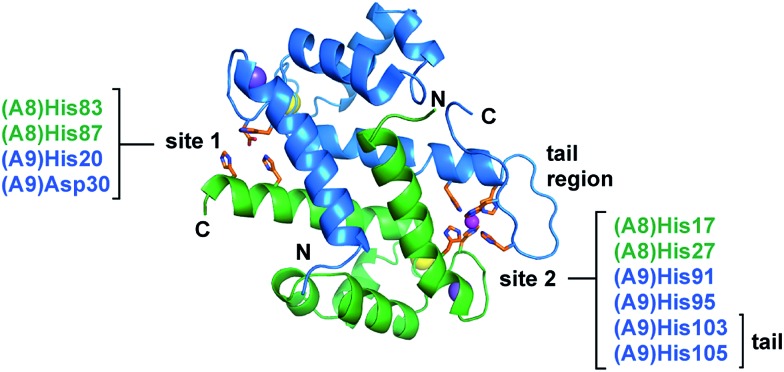
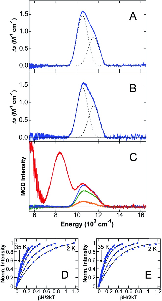
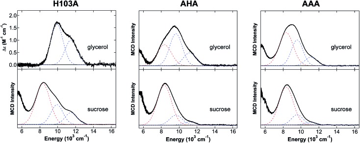
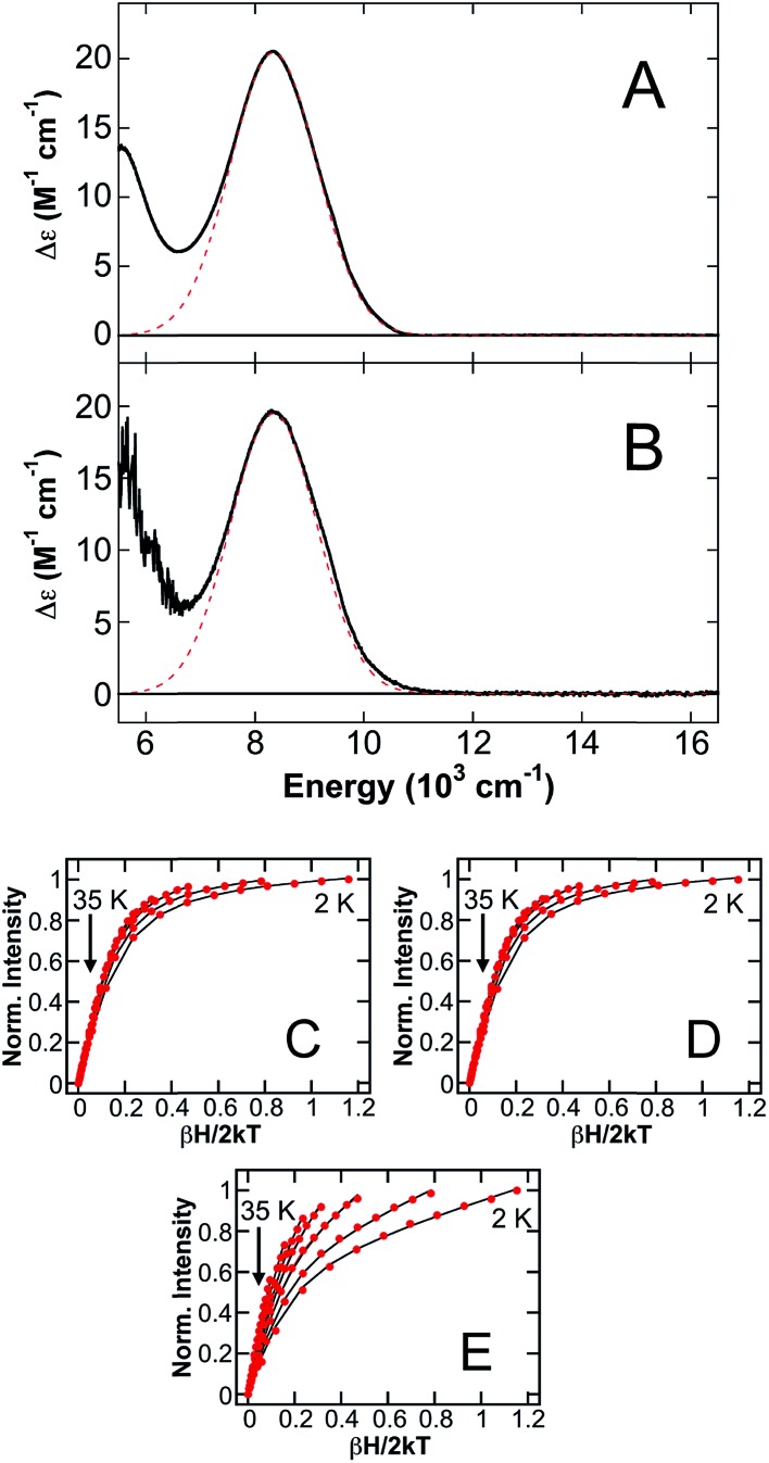
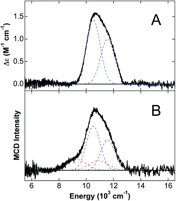
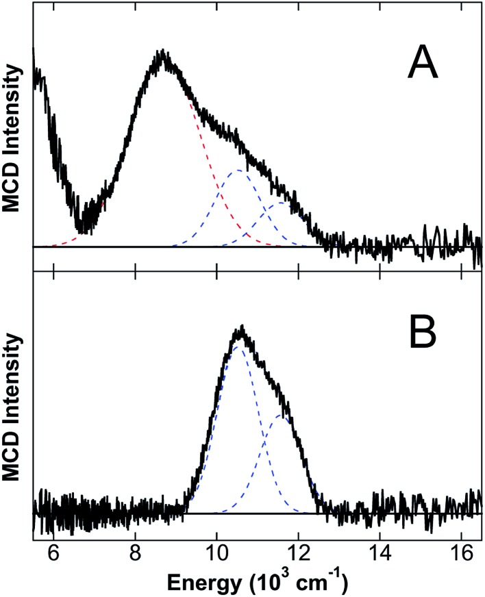
Calprotectin (CP) is an abundant metal-chelating protein involved in host defense, and the ability of human CP to bind Fe(ii) in a calcium-dependent manner was recently discovered. In the present study, near-infrared magnetic circular dichroism spectroscopy is employed to investigate the nature of Fe(ii) coordination at the two transition-metal-binding sites of CP that are a His3Asp motif (site 1) and a His6 motif (site 2). Upon the addition of sub-stoichiometric Fe(ii), a six-coordinate (6C) Fe(ii) center associated with site 2 is preferentially formed in the presence of excess Ca(ii). This site exhibits an exceptionally large ligand field (10Dq = 11 045 cm-1) for a non-heme Fe(ii) protein. Analysis of CP variants lacking residues of the His6 motif supports that CP coordinates Fe(ii) at site 2 by employing six His ligands. In the presence of greater than one equiv. of Fe(ii) or upon mutation of the His6 motif, the metal ion also binds at site 1 of CP to form a five-coordinate (5C) Fe(ii)-His3Asp motif that was previously unidentified in this system. Notably, the introduction of His-to-Ala mutations at the His6 motif results in a mixture of 6C (site 2) and 5C (site 1) signals in the presence of sub-stoichiometric Fe(ii). These results are consistent with a reduced Fe(ii)-binding affinity of site 2 as more weakly coordinating water-derived ligands complete the 6C site. In the absence of Ca(ii), both sites 1 and 2 are occupied upon addition of sub-stoichiometric Fe(ii), and a stronger ligand field is observed for the 5C site. These spectroscopic studies provide further evaluation of a unique non-heme Fe(ii)-His6 site for metalloproteins and support the notion that Ca(ii) ions influence the Fe(ii)-binding properties of CP.


Figures

Fig. 1
Fig. 1. The two transition-metal-binding sites of CP. The Mn(ii)-, Ca(ii)-, and Na(i)-bound CP dimer unit is shown (PDB ; 4XJK). Site 1 comprises His83 and His87 of S100A8 and His20 and Asp30 of S100A9. Site 2 comprises His17 and His27 of S100A8 and His91, His95, His103, and His105 of S100A9. The S100A8 subunit is green, and the S100A9 subunit is blue. Na(i), Ca(ii), and Mn(ii) ions are depicted as purple, yellow, and magenta spheres, respectively. The N- and C-termini of each subunit and the S100A9 C-terminal tail region are labeled.
Fig. 2
Fig. 2. NIR MCD studies of Fe(ii) binding to CP-Ser. The 5 K, 7 T NIR MCD spectra of CP-Ser/Ca(ii)/Fe(ii) in (A) glycerol and (B) sucrose glasses. (C) The 5 K, 7 T NIR MCD spectra for the titration of apo CP-Ser with 0.3 equiv. (orange), 0.6 equiv. (green), 0.9 equiv. (blue) and 1.2 equiv. (red) of Fe(ii). Saturation magnetization data (dots) and best fit (lines) for CP-Ser/Ca(ii)/Fe(ii) in (D) glycerol and (E) sucrose at 10 310 cm–1.
Fig. 3
Fig. 3. NIR MCD studies of site 2 variants of CP-Ser. The 5 K, 7 T NIR MCD spectra of H103A/Ca(ii)/Fe(ii), AHA/Ca(ii)/Fe(ii) and AAA/Ca(ii)/Fe(ii) in glycerol and sucrose. Best fits are shown in dashed lines with blue denoting 6C site LF transitions and red denoting the highest-energy 5C site LF transition (the low energy tail was not fit).
Fig. 4
Fig. 4. NIR MCD studies of ΔHis4. The 5 K, 7 T NIR MCD spectra of ΔHis4/Ca(ii)/Fe(ii) in (A) glycerol and (B) sucrose glasses. Saturation magnetization data (dots) and best fit (lines) for ΔHis4/Ca(ii)/Fe(ii) in glycerol collected at (C) 8905 cm–1, (D) 7810 cm–1 and (E) 5880 cm–1. The low-energy tail transitions were not included in the fits as only part of the transition was observable.
Fig. 5
Fig. 5. NIR MCD studies of ΔHis3Asp. The 5 K, 7 T NIR MCD spectra of (A) ΔHis3Asp/Ca(ii)/Fe(ii) and (B) ΔHis3Asp/Ca(ii) + 1.5 equiv. Fe(ii) in sucrose. Best fits are shown in dashed lines with blue denoting 6C His6 site LF transitions and orange denoting LF transitions due to [Fe(H2O)6]2+.
Fig. 6
Fig. 6. NIR MCD studies of CP-Ser and ΔHis3Asp in the absence of Ca(ii). The 5 K, 7 T NIR MCD spectra of (A) CP-Ser/Fe(ii) and (B) ΔHis3Asp/Fe(ii) in sucrose, where no Ca(ii) is present for either sample. Best fits are shown in dashed lines with blue denoting 6C His6 site LF transitions and red denoting LF transitions due to a 5C Fe(ii) site.

References

    1. Hood M. I., Skaar E. P. Nat. Rev. Microbiol. 2012;10:525–537. - PMC - PubMed
    1. Diaz-Ochoa V. E., Jellbauer S., Klaus S., Raffatellu M. Front. Cell. Infect. Microbiol. 2014;4:2. - PMC - PubMed
    1. Sohnle P. G., Collins-Lech C., Wiessner J. H. J. Infect. Dis. 1991;164:137–142. - PubMed
    1. Corbin B. D., Seeley E. H., Raab A., Feldmann J., Miller M. R., Torres V. J., Anderson K. L., Dattilo B. M., Dunman P. M., Gerads R., Caprioli R. M., Nacken W., Chazin W. J., Skaar E. P. Science. 2008;319:962–965. - PubMed
    1. Clohessy P. A., Golden B. E. Scand. J. Immunol. 1995;42:551–556. - PubMed

LinkOut - more resources