. 2017 Apr 1;8(4):3038-3046.
doi: 10.1039/c6sc04127b. Epub 2017 Feb 3.

Plasmonic labeling of subcellular compartments in cancer cells: multiplexing with fine-tuned gold and silver nanoshells

Affiliations

Plasmonic labeling of subcellular compartments in cancer cells: multiplexing with fine-tuned gold and silver nanoshells

R G Sobral-Filho et al. Chem Sci. .

Abstract

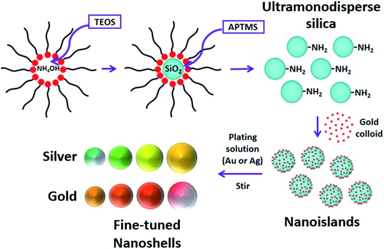
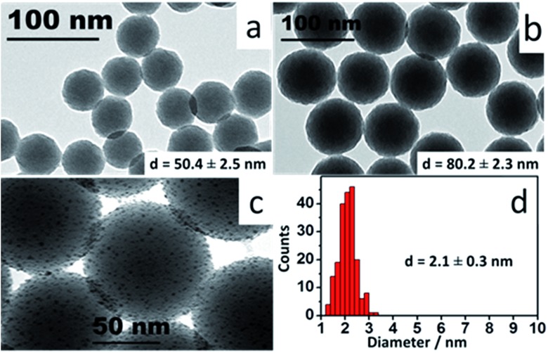
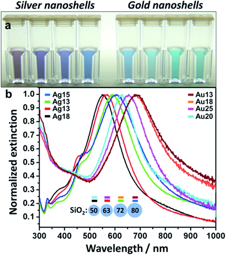
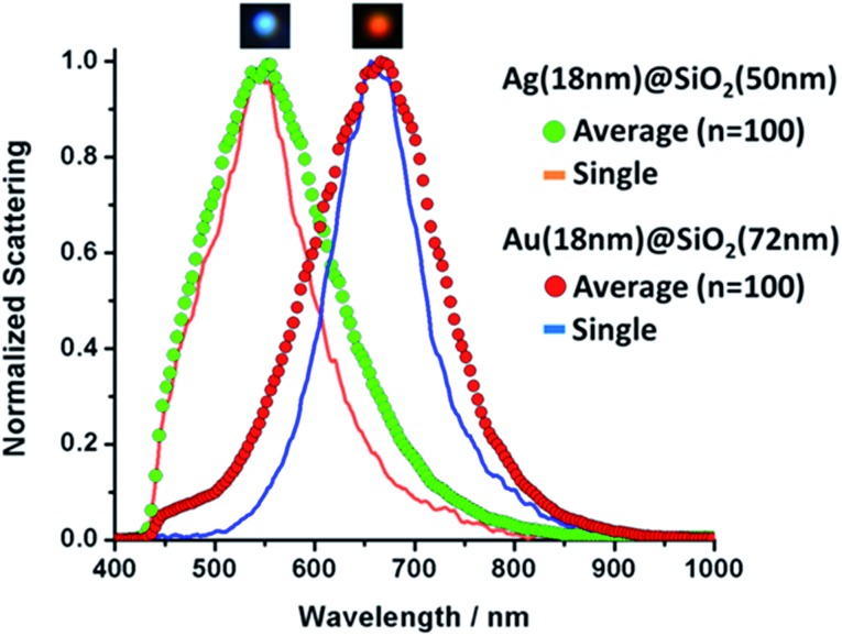
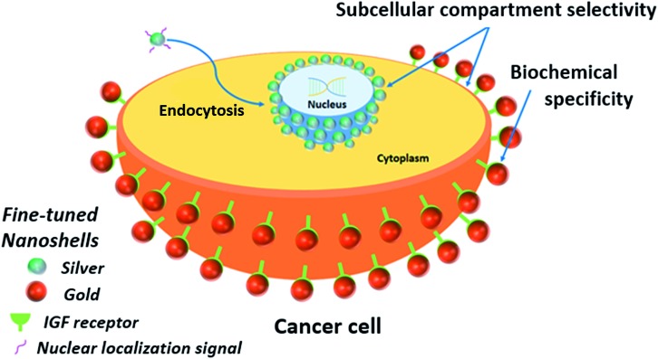
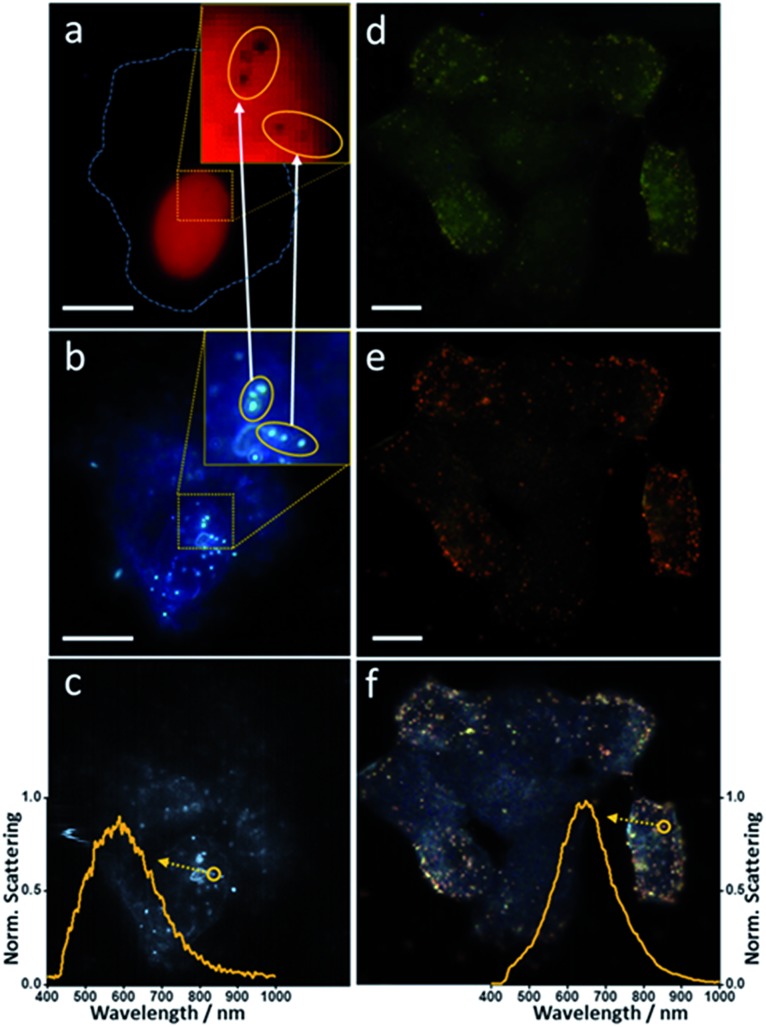
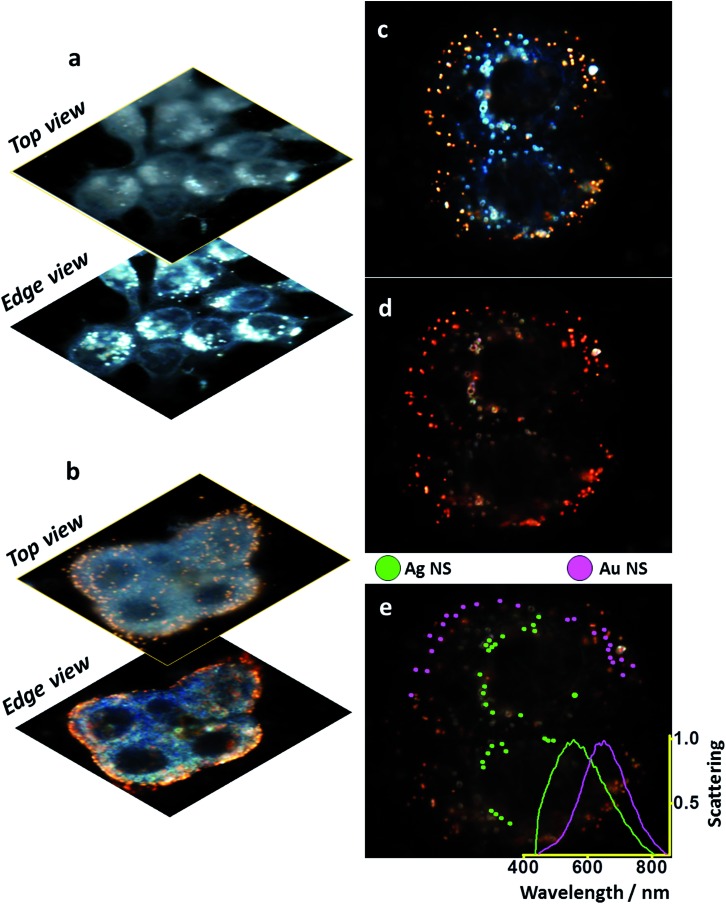
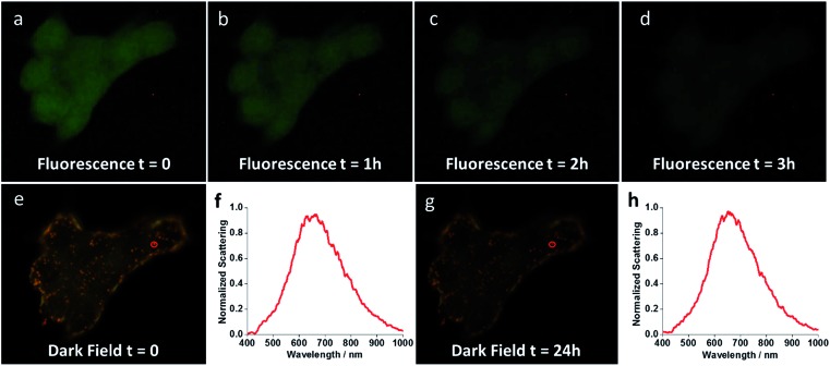
Fine-tuned gold and silver nanoshells were produced via an entirely reformulated synthesis. The new method yielded ultramonodisperse samples, with polydispersity indexes (PI) as low as 0.02 and narrow extinction bands suited for multiplex analysis. A library of nanoshell samples with localized surface plasmon resonances (LSPR) spanning across the visible range was synthesized. Hyperspectral analysis revealed that the average scattering spectrum of 100 nanoshells matched closely to the spectrum of a single nanoshell, indicating an unprecedented low level of nanoparticle-to-nanoparticle variation for this type of system. A cell labeling experiment, targeting different subcellular compartments in MCF-7 human breast cancer cells, demonstrated that these monodisperse nanoparticles can be used as a multiplex platform for single cell analysis at the intracellular and extracellular level. Antibody-coated gold nanoshells targeted the plasma membrane, while silver nanoshells coated with a nuclear localization signal (NLS) targeted the nuclear membrane. A fluorescence counterstaining experiment, as well as single cell hyperspectral microscopy showed the excellent selectivity and specificity of each type of nanoparticle for its designed subcellular compartment. A time-lapse photodegradation experiment confirmed the enhanced stability of the nanoshells over fluorescent labeling and their capabilities for long-term live cell imaging.


Figures

Scheme 1
Scheme 1. Ultramonodisperse aminated silica particles are produced in a one-batch synthesis via a reverse microemulsion system. Small gold nanoparticles are then attached to the silica and the shell growth takes place under stirring in a plating solution with metal ions at low concentration (150 μM). Different SiO2 sizes and shell thicknesses can be achieved with this method.
Fig. 1
Fig. 1. TEM images: (a and b) SiO2 nanoparticles synthesized by reverse microemulsion; (c) nanoislands at high magnification; (d) Au colloid diameter histogram.
Fig. 2
Fig. 2. (a) Fine-tuned Ag and Au nanoshells samples – cuvettes match measured spectra in 3b (left to right); (b) extinction spectra for Ag and Au nanoshells – core size and shell thickness are color-coded to the curves (dimensions in nm).
Fig. 3
Fig. 3. Single and averaged (n = 100) scattering spectra for Ag and Au nanoshells. Inserts above each curve show individual nanoshells as seen under dark field illumination.
Scheme 2
Scheme 2. Labeling of subcellular compartments by ultramonodisperse nanoshells. Silver nanoshells bioconjugated to a nuclear localization signal (NLS) are internalized by the cells. The NLS peptide leads the particles to escape nonspecific endosomal and exocytic pathways and accumulate on the nuclear membrane. Gold nanoshells bioconjugated to an anti-IGFR antibody, target the insulin receptors (IGFR) localized on the plasma membrane of MCF-7 cells. Selectivity of the nanoshells for each subcellular compartment is evidenced, as well as the ability to target specific biochemical elements within such compartments, as showed for the insulin receptors and the antibody-coated gold nanoshells.
Fig. 4
Fig. 4. Combined microscopical (fluorescence, dark field and hyperspectral) analysis of nanoshell-labeling. (a) and (d) – fluorescence microscopy, (b) and (e) – conventional white light dark field microscopy, (c) and (f) – hyperspectral dark field microscopy. Inserts in (a) and (b) show magnified regions of interest on the nuclear membrane. Inserts in (c) and (f) evidence the scattering spectra of single silver (c) and gold (f) nanoshells. Cell membrane is outlined by dashed line in (a). Scale bar 10 μm. Measurement details in the ESI file.
Fig. 5
Fig. 5. Multiplex-imaging different MCF-7 cell compartments with Ag and Au nanoshells. Different focal depths for: (a) Ag-NLS – targeting the nuclear membrane (b) Au-IGFR – targeting the plasma membrane; same field: (c) regular dark field image, (d) hyperspectral dark field image, (e) hyperspectral sorting between Ag and Au nanoshells – Ag (18 nm)@SiO2 (50 nm) labeling the nuclear membrane and Au (18 nm)@SiO2 (72 nm) labeling IGF receptors on the plasma membrane, insert shows averaged scattering spectra for the selected nanoshells.
Fig. 6
Fig. 6. Long-term photostability of nanoshells. Time-lapse images show fluorescent phalloidin photodegrading over the course of a few hours under illumination (a–d), whereas Au-IGFR nanoshells remain active even after 24 h under illumination (e and g). (f and h) show the single scattering spectrum of the same gold nanoshell (red circular inserts in e and g) at t = 0 and t = 24 h.

Similar articles

Cited by

References

    1. Chou L. Y., Zagorovsky K., Chan W. C. Nat. Nanotechnol. 2014;9:148–155. - PMC - PubMed
    1. Brito-Silva A. M., de Araujo C. B., Brayner F. A., Santos S. S., Galembeck A., Milet E. R. Polym. Eng. Sci. 2010;50:2350–2355.
    1. Brito-Silva A. M., Sobral-Filho R. G., Mejía H. A., Wang Y.-H., Wang P., Machado G., Falcão-Filho E. L., de Araújo C. B., Brolo A. G. J. Phys. Chem. C. 2014;118:18372–18376.
    1. Bardhan R., Lal S., Joshi A., Halas N. J. Acc. Chem. Res. 2011;44:936–946. - PMC - PubMed
    1. Choi Y., Park Y., Kang T., Lee L. P. Nat. Nanotechnol. 2009;4:742–746. - PubMed