. 2017 Apr 1;8(4):3080-3091.
doi: 10.1039/c6sc05420j. Epub 2017 Jan 30.

DNA-barcoded labeling probes for highly multiplexed Exchange-PAINT imaging

Affiliations

DNA-barcoded labeling probes for highly multiplexed Exchange-PAINT imaging

Sarit S Agasti et al. Chem Sci. .

Abstract

Recent advances in super-resolution fluorescence imaging allow researchers to overcome the classical diffraction limit of light, and are already starting to make an impact in biology. However, a key challenge for traditional super-resolution methods is their limited multiplexing capability, which prevents a systematic understanding of multi-protein interactions on the nanoscale. Exchange-PAINT, a recently developed DNA-based multiplexing approach, in theory facilitates spectrally-unlimited multiplexing by sequentially imaging target molecules using orthogonal dye-labeled 'imager' strands. While this approach holds great promise for the bioimaging community, its widespread application has been hampered by the availability of DNA-conjugated ligands for protein labeling. Herein, we report a universal approach for the creation of DNA-barcoded labeling probes for highly multiplexed Exchange-PAINT imaging, using a variety of affinity reagents such as primary and secondary antibodies, nanobodies, and small molecule binders. Furthermore, we extend the availability of orthogonal imager strands for Exchange-PAINT to over 50 and assay their orthogonality in a novel DNA origami-based crosstalk assay. Using our optimized conjugation and labeling strategies, we demonstrate nine-color super-resolution imaging in situ in fixed cells.


Figures

Fig. 1
Fig. 1. Crosstalk experiment to check the orthogonality of 52 docking sequences. (a) DNA origami carries single-stranded extensions (docking strands), which can transiently bind fluorescently labeled oligonucleotides (imagers) in solution. (b) Rectangular origami with modified extended staples (left side); a schematic representation of the structure is located on the right side; each hexagon represents a staple position that can be extended for DNA-PAINT imaging. Each origami contains a unique 6-bit barcode, addressable with the sequence P1 (left side), and single-stranded extensions that will act as docking sites for the imagers to be tested (P2–P52). Together, these extensions form a mirrored “F” shape (right side). (c) Crosstalk check for sequence P40. The upper row shows schematic representations of the barcode structures for each sequence. The bottom row shows the experimental data. The mirrored “F” appears only next to the barcode for the P40 sequence. This shows the orthogonality of the P40 sequence to all other sequences. (d) Overview image of the crosstalk experiment for P40. Scale bars: 50 nm (c), 200 nm (d).
Fig. 2
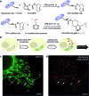
Fig. 2. Antibody-DNA conjugation method and super-resolution imaging with a DNA-conjugated secondary antibody. (a) Synthesis scheme for DNA-conjugated antibody preparation. Note that SM(PEG)2 is depicted here as NHS-EG2-Mal. (b) Labeling strategy for the DNA-conjugated secondary antibody. (c) Secondary antibody-based DNA-PAINT super-resolution imaging of microtubules inside a fixed BSC-1 cell. Zooming in of the highlighted area shows the resolution improvement compared to the diffraction-limited micrographs of the same area. The cross-sectional histogram of a hollow microtubule structure clearly shows two distinct lines with a separation of ∼40 nm. This is in good agreement with earlier reports.
Fig. 3
Fig. 3. DNA-PAINT imaging with a DNA-conjugated primary antibody. (a) Labeling scheme with a DNA-conjugated primary antibody. (b) Primary antibody-based DNA-PAINT imaging of microtubules inside a fixed BSC-1 cell. (c) Primary antibody-based DNA-PAINT imaging of Tom20 in mitochondria. Tom20 localizes to the mitochondrial membrane, which is clearly resolved. Scale bars: 5 μm.
Fig. 4
Fig. 4. Synthesis of DNA-conjugated nanobodies for DNA-PAINT imaging. (a) Synthesis scheme for DNA-conjugated nanobody preparation. (b) Labeling scheme using the DNA-conjugated nanobody. (c, d) Nanobody-based DNA-PAINT super-resolution imaging of the mitochondrial network inside a fixed HeLa cell. A comparison of the diffraction-limited image (c) to the DNA-PAINT image (d) underlines the achieved resolution increase. Scale bars: 5 μm.
Fig. 5
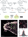
Fig. 5. Conjugation of DNA oligos to phalloidin for actin imaging with DNA-PAINT. (a) Synthesis scheme for DNA-conjugated phalloidin. (b) Labeling strategy for phalloidin using the DNA-phalloidin conjugate. (c) Resulting DNA-PAINT image of the actin network inside a fixed HeLa cell. Zooming in to the highlighted area (green) highlights the achievable resolution. A Gaussian distribution was fitted to the cross-sectional histogram of an actin fiber (selected from the highlighted red region). FWHM of the distribution: ∼12 nm.
Fig. 6
Fig. 6. Secondary antibody-based labeling for multiplexing with Exchange-PAINT. (a) Schematic representation of Exchange-PAINT. Target proteins (T1···T8) are labeled with DNA (D1···D8)-conjugated secondary antibodies using an indirect immunostaining approach. Complementary ATTO655-dye-labeled DNA strands (I1···I8) are sequentially applied to the sample. Post-acquisition, a washing buffer with reduced ionic strength was used to efficiently remove the imagers. Eight imaging rounds were performed using orthogonal imager strands with the same dye. (b) Eight-target DNA-PAINT image of fixed HeLa cells acquired in eight sequential rounds. Scale bars: 5 μm.
Fig. 7
Fig. 7. Primary antibody-based labeling for multiplexing with Exchange-PAINT. (a) Labeling strategy for primary antibody-based imaging. The target proteins (T1···Tn) were labeled with DNA (D1···Dn)-conjugated primary antibodies using a direct immunostaining approach. Complementary imager strands (labeled with ATTO655) were sequentially introduced to the sample for super-resolution imaging as before. Post-acquisition, a washing buffer with reduced ionic strength was introduced to remove all imagers. Nine imaging rounds were performed using orthogonal imager strands conjugated to the same dye. (b) Nine-target super-resolution image of proteins in fixed HeLa cells acquired using nine rounds of Exchange-PAINT.

References

    1. Knoll M., Ruska E. Z. Phys. 1932;78:318–339.
    1. Coons A. H., Creech H. J., Jones R. N. Exp. Biol. Med. 1941;47:200–202.
    1. Hell S. W. J. Phys. D: Appl. Phys. 2015;48:443001.
    1. Gustafsson M. G. J. Microsc. 2000;198:82–87. - PubMed
    1. Hell S. W., Wichmann J. Opt. Lett. 1994;19:780–782. - PubMed