Review
. 2017 Apr 13:4:21.
doi: 10.3389/fmolb.2017.00021. eCollection 2017.

The Interplay of Cofactor Interactions and Post-translational Modifications in the Regulation of the AAA+ ATPase p97

Affiliations
Review

The Interplay of Cofactor Interactions and Post-translational Modifications in the Regulation of the AAA+ ATPase p97

Petra Hänzelmann et al. Front Mol Biosci. .

Abstract

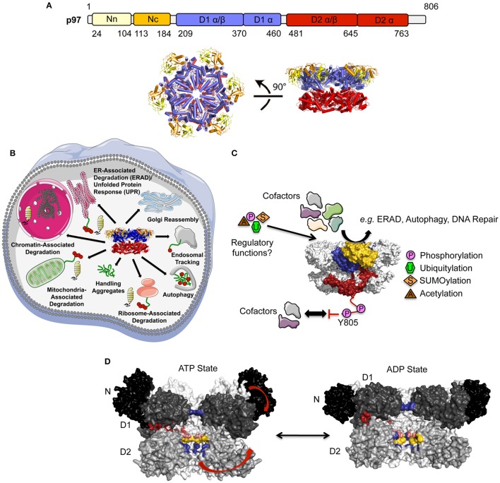
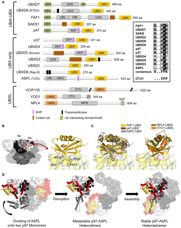
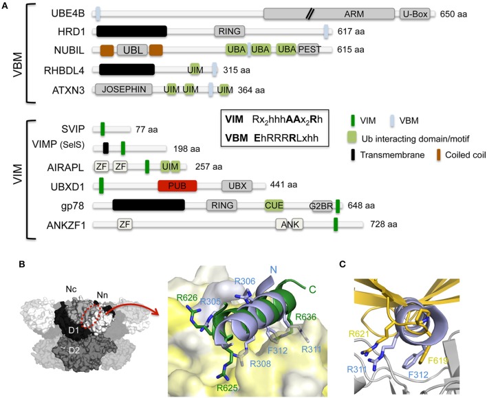
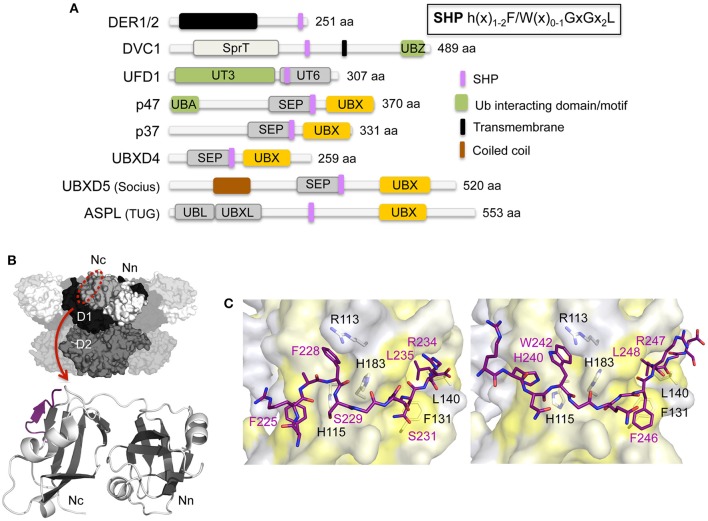
The hexameric type II AAA ATPase (ATPase associated with various activities) p97 (also referred to as VCP, Cdc48, and Ter94) is critically involved in a variety of cellular activities including pathways such as DNA replication and repair which both involve chromatin remodeling, and is a key player in various protein quality control pathways mediated by the ubiquitin proteasome system as well as autophagy. Correspondingly, p97 has been linked to various pathophysiological states including cancer, neurodegeneration, and premature aging. p97 encompasses an N-terminal domain, two highly conserved ATPase domains and an unstructured C-terminal tail. This enzyme hydrolyzes ATP and utilizes the resulting energy to extract or disassemble protein targets modified with ubiquitin from stable protein assemblies, chromatin and membranes. p97 participates in highly diverse cellular processes and hence its activity is tightly controlled. This is achieved by multiple regulatory cofactors, which either associate with the N-terminal domain or interact with the extreme C-terminus via distinct binding elements and target p97 to specific cellular pathways, sometimes requiring the simultaneous association with more than one cofactor. Most cofactors are recruited to p97 through conserved binding motifs/domains and assist in substrate recognition or processing by providing additional molecular properties. A tight control of p97 cofactor specificity and diversity as well as the assembly of higher-order p97-cofactor complexes is accomplished by various regulatory mechanisms, which include bipartite binding, binding site competition, changes in oligomeric assemblies, and nucleotide-induced conformational changes. Furthermore, post-translational modifications (PTMs) like acetylation, palmitoylation, phosphorylation, SUMOylation, and ubiquitylation of p97 have been reported which further modulate its diverse molecular activities. In this review, we will describe the molecular basis of p97-cofactor specificity/diversity and will discuss how PTMs can modulate p97-cofactor interactions and affect the physiological and patho-physiological functions of p97.

Keywords: AAA+ ATPase; cofactor diversity; conformational changes; p97; post-translational modification; protein disassembly; protein quality control.


Figures

Figure 1
Figure 1
Structure and function of p97. (A) Top, Domain architecture of p97. The N domain can be further subdivided into an N- (Nn, colored in yellow) and a C-terminal (Nc, colored in orange) subdomain, the two ATPase domains (colored in blue and red, respectively) each feature a larger α/β and a smaller α-helical subdomain. Bottom, top (left) and side views (right) of the p97 hexamer (pdb entry 3CF1; Davies et al., 2008) (adapted from Buchberger et al., 2015). (B) Cellular functions of p97. (C) Regulation of p97 function by cofactor interactions and post-translational modifications (PTMs). p97 is colored as in (A) for one subunit and gray for the remaining subunits. (D) Conformational changes of p97 upon ATP binding/hydrolysis. Molecular surface of the p97 central cavity (side view) in the ATP- and ADP-bound states (pdb entries 5FTN and 5FTK; Banerjee et al., 2016) with two monomers in black/dark/medium (N/D1/D2) gray and two monomers in light gray. For clarity, the two monomers in the front are not shown. The restriction in the D1 domain (His-gate) is shown in blue. Catalytically important positively charged (Arg586 and Arg599, colored in blue), negatively charged (Glu544, colored in pink), and hydrophobic (Trp551 and Phe552, colored in yellow) residues lining the D2 pore are indicated. Conformational changes are indicated with arrows and the opening at the D1D2 interface is shown in red.
Figure 2
Figure 2
Interaction of p97 with cofactors containing UBX and UBXL domains. (A) Domain architecture of mammalian UBX and UBXL domain cofactors. A sequence alignment of the R…FPR motif in the UBX domains is shown on the right. MPN, (Mpr1, Pad1 N-terminal); NZF, NPL4 zinc finger; OTU, ovarian tumor; SEP, Shp, eyes-closed, p47; SHP, binding segment 1 (BS1); UAS, domain of unknown function found in FAF1 and other proteins; UBA, ubiquitin-associated; UBL, ubiquitin-like; UBX, ubiquitin regulatory X; UBXL, UBX-like; UIM, ubiquitin-interacting motif; ZF, zinc finger. (B) Left, molecular surface of p97 [N domain in white (Nn)/light gray (Nc), D1 in black, D2 in dark gray] with the UBX/UBXL binding site indicated. Right, FAF1-UBX—p97 N complex (pdb entry 3QQ8; Hänzelmann et al., 2011). The UBX domain (colored in gold) is shown in cartoon representation and p97 N as molecular surface (colored in shades of yellow according to hydrophobicity). The R…FPR motif is shown in stick representation. (C) Left, superposition of the UBX domains from FAF1 (pdb entry 3QQ8, colored in gold; Hänzelmann et al., 2011), p47 (pdb entry 1S3S, colored in brown; Dreveny et al., 2004) and ASPL (pdb entry 5IFW, colored in gray; Arumughan et al., 2016) bound to p97 N colored as in (B). Right, superposition of the UBXL domains of NPL4 (pdb entry 4RV0, colored in olive; Hao et al., 2015) and OTU1 (pdb entry 4KDI, colored in orange; Kim et al., 2014) bound to p97 N colored as in (B). (D) Disassembly of p97 hexamers through the interaction with the ASPL-UBX domain and formation of the stable p97-ASPL heterotetramer via metastable p97-ASPL heterodimers. For clarity, only two monomers of p97 are shown. One heterodimer is shown in cartoon representation and the other in surface representation. The ASPL-UBX domain is colored in yellow with the N- and C-terminal extensions in red and p97 in light gray (N domain), dark gray (D1 domain) and gray (D2 domain) (pdb entry 5IFW; Arumughan et al., 2016). The curved arrow indicates the reorientation of the D2 domain.
Figure 3
Figure 3
Interaction of p97 with cofactors harboring either a VBM or VIM binding motif. (A) Domain architecture of mammalian cofactors with a VBM or VIM binding motif. The consensus sequences for both binding motifs are shown in the inset. ANK, ankyrin repeat; ARM, armadillo/beta-catenin-like repeats; CUE, coupling of ubiquitin conjugation to endoplasmic reticulum degradation; G2BR, Ube2g2-binding region; JOSEPHIN, deubiquitinase domain; PEST, PEST motif, Pro, Glu, Ser and Thr rich sequence; PUB, PNGase/UBA or UBX containing proteins; RING, RING (Really Interesting New Gene) finger; U-Box, UFD2-homology domain; UBA, ubiquitin-associated; UBL, ubiquitin-like; UIM, ubiquitin-interacting motif; VBM, VCP-binding motif; VIM, VCP-interacting motif; ZF, zinc finger. (B) Left, molecular surface of p97 [N domain in white (Nn)/light gray (Nc), D1 in black, D2 in dark gray] with the VIM/VBM binding site indicated. Right, cartoon representation of a superposition of the gp78 VIM (pdb entry 3TIW, colored in green; Hänzelmann and Schindelin, 2011) and RHBDL4 VBM (pdb entry 5EPP, colored in light blue; Lim et al., 2016a) binding motifs in complex with the p97 N domain (molecular surface colored according to hydrophobicity). Key interactions are shown in stick representation. (C) Cartoon representation of a superposition of the RHBDL4 VBM (pdb entry 5EPP, colored in light blue; Lim et al., 2016a) and the FAF1-UBX (pdb entry 3QQ8, colored in gold; Hänzelmann et al., 2011) p97 N (colored in light gray) complexes. The side chains of the RF dipeptide of the VBM and the FPR motif of the VIM are shown in stick representation.
Figure 4
Figure 4
Interaction of p97 with cofactors harboring a SHP binding motif. (A) Domain architecture of mammalian cofactors with a SHP binding motif. The consensus sequence for the SHP binding motif is shown in the inset. SEP, Shp, eyes-closed, p47; SHP, binding segment 1 (BS1); SprT, SprT-like; UT3 and UT6, UFD1 domains; UBA, ubiquitin-associated; UBL, ubiquitin-like; UBX, ubiquitin regulatory X; UBXL, UBX-like; UBZ, ubiquitin-binding zinc finger; UIM, ubiquitin-interacting motif; ZF, zinc finger. (B) Top, molecular surface of p97 [N domain in white (Nn)/light gray (Nc), D1 in black, D2 in dark gray] with the SHP binding site indicated. Bottom, cartoon representation of the overall structure of the UFD1 SHP binding motif (colored in purple) bound to the p97 N domain (β-strands in dark gray and α-helices in light gray) (pdb entry 5B6C; Le et al., 2016). (C) Stick representations of the SHP binding motifs of UFD1 (left, pdb entry 5B6C, colored in purple; Le et al., 2016) and DER1 (right, pdb entry 5GLF, colored in purple; Lim et al., 2016b). The p97 N domain is shown as molecular surface (colored according to hydrophobicity). Key interactions are shown.
Figure 5
Figure 5
Interaction of p97 with PUB and PUL domain-containing cofactors. (A) Domain architecture of mammalian PUB and PUL domain-containing cofactors. NZF, NPL4 zinc finger; PAW, present in PNGases and other worm proteins; PFU, PLAA family ubiquitin binding domain; PUB, PNGase/UBA or UBX containing proteins; PUL, (PLAP, Ufd3, and Lub1p); RING, RING (Really Interesting New Gene) finger; TGc, transglutaminase like; UBA, ubiquitin-associated; UBX, ubiquitin regulatory X; WD40, WD40 β-propeller. (B) Left top, molecular surface of p97 [N domain in white (Nn)/light gray (Nc), D1 in black, D2 in dark gray] with the p97 C-terminal cofactor binding site indicated. Left bottom, overall structure of the p97 C-terminus (stick representation) bound to the PNGase PUB domain shown in cartoon representation (pdb entry 2HPL; Zhao et al., 2007) (adapted from Buchberger et al., 2015). Right, stick representation of the p97 C-terminus together with the molecular surface of the PNGase (top, pdb entry 2HPL, Zhao et al., 2007) and the HOIP (bottom, pdb entry 4P0A; Schaeffer et al., 2014) PUB domains (colored according to hydrophobicity). Key interactions are shown. (C) Top, overall structure of the p97 C-terminus (stick representation) bound to the PLAA PUL domain shown in cartoon representation (pdb entry 3EEB; Qiu et al., 2010) (modified from Buchberger et al., 2015). Bottom, stick representation of the p97 C-terminus together with the molecular surface of the PLAA PUL domain (colored according to hydrophobicity). The arrow indicates a possible location of the N-terminal extension of the p97 C-terminal residues. Key interactions are shown.
Figure 6
Figure 6
Regulation of p97 cofactor binding to the N domain. (A) Binding site competition. Superposition of p97 N domain cofactor complexes: SHP of UFD1 (pdb entry 5B6C, colored in purple; Le et al., 2016), VIM of gp78 (pdb entry 3TIW, colored in green; Hänzelmann and Schindelin, 2011), VBM of RHBDL4 (pdb entry 5EPP, colored in light blue; Lim et al., 2016b), FAF1-UBX (pdb entry 3QQ8, colored in gold; Hänzelmann et al., 2011) and NPL4-UBXL (pdb entry 4RV0, colored in olive; Hao et al., 2015). Cofactors are shown in cartoon or stick representation and p97 N as molecular surface (Nn in dark gray, Nc in light gray). (B) Conformational changes of p97 upon ATP binding/hydrolysis. Side view of the molecular surface of p97 in the ATP- and ADP-bound states (pdb entries 5FTN and 5FTK; Banerjee et al., 2016) together with a cartoon representation of the FAF1-UBX domain (pdb entry 3QQ8, colored in gold; Hänzelmann et al., 2011) and the gp78 VIM (pdb entry 3TIW, colored in green; Hänzelmann and Schindelin, 2011) as well as a stick representation of the UFD1 SHP binding motif (pdb entry 5B6C, colored in purple; Le et al., 2016). (C) Models for bipartite binding to p97. UFD1-NPL4 and p47 interact with either identical or different N domains (adapted with permission from Hänzelmann and Schindelin, 2016a) whereas UBXD1 simultaneously binds to the N domain and the unstructured C-terminus (C-T) (modified from Buchberger et al., 2015). p97 is shown in a cartoon representation with the N domains colored in yellow. (D) Models for different oligomeric assemblies and higher order complexes for different cofactors as indicated. p97 is shown in a cartoon representation with the N domains colored in yellow.
Figure 7
Figure 7
p97 post-translational modifications (PTMs). (A) Domain architecture of p97 together with identities of PTMs derived from the public database PhosphoSitePlus (Hornbeck et al., 2015) and published sources. (B) Nucleotide-induced conformational changes of the p97 N-terminal extension (residues 1–24, colored black). Molecular surface representation of p97 in the ADP and ATP states (pdb entries 5FTK and 5FTN; Banerjee et al., 2016). In one of the monomers of the ATP-bound structure the N-terminal extension is modeled according to Schuller et al. (2016). The opening at the D1D2 interface is shown in red. Identified PTMs on the extension are indicated. (C) Nucleotide-induced conformational changes of the p97 C-terminus. Molecular surface representation together with a cartoon representation of the C-terminus in the apo- (top) and ATP-bound state (bottom) (pdb entries 5C19 and 5C18; Hänzelmann and Schindelin, 2016b). The disordered C-terminal tail is indicated with dashed lines. Identified PTMs in the C-terminal helix α9 are shown and listed for the disordered region. (D) Phosphorylation sites identified in the ND1 part of p97 (N-terminal extension in black, N domain in dark gray, D1 in light gray). Identified phosphorylation sites (colored in magenta) are mapped onto the molecular surface of p97 in the ADP- and ATP-bound states (pdb entries 5FTK and 5FTN; Banerjee et al., 2016) and are shown in a side view and top view. Residues in the D1D2 interface are shown in red. In addition, palmitoylation of Cys105 (colored in yellow) and monomethylation of Arg155 (colored in blue) are indicated. (E) Ubiquitylation and SUMOylation sites identified on the ND1 part of p97. Identified ubiquitylation sites (colored in green) and SUMOylation sites (colored in orange) as well as sites, which carry both modifications (colored in cyan), are mapped onto the molecular surface of p97 as in (D). p97 is colored as in (D).
Figure 8
Figure 8
Models of p97 disassembly activity. (A) Proposed p97 translocation pathways. Molecular surface of the p97 central cavity (side view) in the ATP bound state (pdb entry 5FTN; Banerjee et al., 2016) with two monomers in black/dark/medium (N/D1/D2) gray and two monomers in light gray. For clarity, the two monomers in the front are not shown. The restriction in the D1 domain (His-gate) is shown in blue, the two D2 pore loops are colored in yellow and forest, respectively and the opening at the D1D2 interface is shown in red. Proposed translocation pathways according to the different models (threading, side access and D2 in-out) are indicated with red arrows. (B) Models for the disassembly function of p97 (translocation-independent model). In the ATP-bound state the N domains are in the up-position and interact via bound cofactors for example with a ubiquitylated substrate being part of a chromatin-associated protein complex. Upon ATP-hydrolysis the down-movement of the N domain would exert a force on the bound substrate, which would disassemble it from the protein complex (left). Depending on the substrate, alternatively the down-movement of the N domain would bring the cofactor bound substrate to the D1D2 interface and unfolded regions of the substrate could enter through this path into the p97 D2 pore with its putative substrate binding loops, which could provide an additional pulling force (right, hybrid model).

References

    1. Akutsu M., Dikic I., Bremm A. (2016). Ubiquitin chain diversity at a glance. J. Cell Sci. 129, 875–880. 10.1242/jcs.183954 - DOI - PubMed
    1. Alexandru G., Graumann J., Smith G. T., Kolawa N. J., Fang R., Deshaies R. J. (2008). UBXD7 binds multiple ubiquitin ligases and implicates p97 in HIF1alpha turnover. Cell 134, 804–816. 10.1016/j.cell.2008.06.048 - DOI - PMC - PubMed
    1. Almeida B., Abreu I. A., Matos C. A., Fraga J. S., Fernandes S., Macedo M. G., et al. (2015). SUMOylation of the brain-predominant Ataxin-3 isoform modulates its interaction with p97. Biochim. Biophys. Acta 1852, 1950–1959. 10.1016/j.bbadis.2015.06.010 - DOI - PubMed
    1. Arumughan A., Roske Y., Barth C., Forero L. L., Bravo-Rodriguez K., Redel A., et al. (2016). Quantitative interaction mapping reveals an extended UBX domain in ASPL that disrupts functional p97 hexamers. Nat. Commun. 7:13047. 10.1038/ncomms13047 - DOI - PMC - PubMed
    1. Ballar P., Shen Y., Yang H., Fang S. (2006). The role of a novel p97/valosin-containing protein-interacting motif of gp78 in endoplasmic reticulum-associated degradation. J. Biol. Chem. 281, 35359–35368. 10.1074/jbc.M603355200 - DOI - PubMed

LinkOut - more resources