. 2017 Mar 17;5(3):2325967117696281.
doi: 10.1177/2325967117696281. eCollection 2017 Mar.

Open Reduction Internal Fixation of Isolated Chondral Fragments Without Osseous Attachment in the Knee: A Case Series

Affiliations

Open Reduction Internal Fixation of Isolated Chondral Fragments Without Osseous Attachment in the Knee: A Case Series

Patrick N Siparsky et al. Orthop J Sports Med. .

Abstract

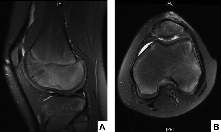
Background: Isolated chondral fractures of the knee are a rare and challenging problem, typically occurring with an acute traumatic event such as dislocation of the patella or ligamentous injury. Historically, repair of unstable chondral fragments without osseous attachment has not been recommended due to concerns about the limited healing potential of cartilage.

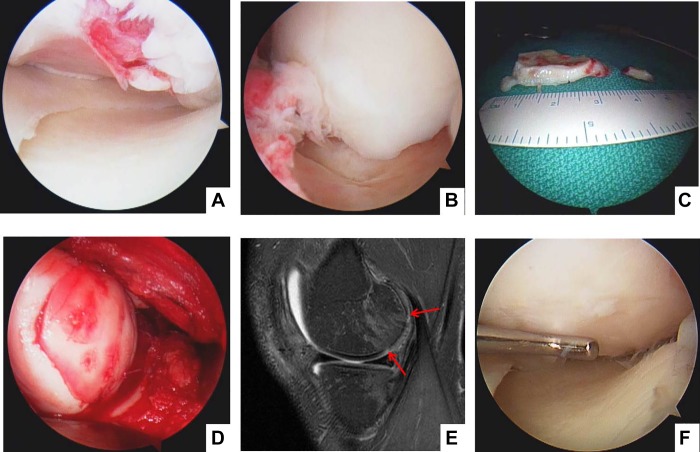
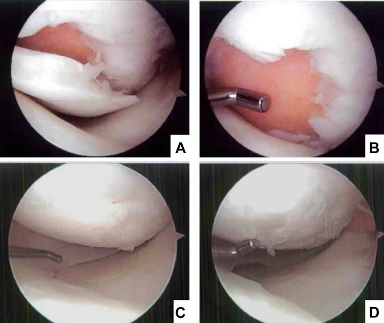
Purpose: To describe a technique for fixation of large isolated chondral fractures of the knee and present 3 cases where large chondral fragments without osseous attachment were fixed successfully with chondral darts and biologic adhesive.

Study design: Case series; Level of evidence, 4.

Methods: The senior author reviewed his case logs for all patients on whom he performed open reduction and internal fixation on large isolated cartilage fragments without osseous attachment. Three were extracted from his review. The clinical and radiographic outcomes were retrospectively reviewed.

Results: Successful results and complete healing was obtained in all 3 patients. This procedure can be done in the setting of concurrent injury, such as anterior cruciate ligament tear, using single- or multistaged chondral repair and ligament reconstruction techniques.

Conclusion: Isolated chondral fragment repair techniques provide the orthopaedic surgeon an additional option for treating these challenging injuries. Primary fixation can be accomplished for what have been historically considered "unsalvageable" fragments.

Keywords: cartilage; chondral; chondral fracture; fixation.


Conflict of interest statement

The authors declared that they have no conflicts of interest in the authorship and publication of this contribution.

Figures

Figure 1.
Figure 1.
A 13-year-old male with large lateral femoral condyle chondral fracture after patellar dislocation. (A) T2-weighted sagittal image with a large chondral fragment (arrow) resting in the suprapatellar pouch. (B) T2-weighted sagittal image showing a lateral femoral condyle chondral defect (edges framed by arrows). (C) T2-weighted axial image with a large lateral femoral condyle defect (edges framed by arrows).
Figure 2.
Figure 2.
Chondral defect preparation. (A) A medial parapatellar approach allows patellar eversion. The defect is exposed and fibrinous material removed. (B) Curette was used to debride the calcified cartilage layer. (C) Free chondral fragment trimmed to fit back in the defect. The fragment edges should be sharply cleaned with a No. 15 blade.
Figure 3.
Figure 3.
Chondral fragment fixation and sealing. (A) The fragment is fixed with chondral darts, evenly spaced, supporting the edges to avoid tilting. (B) Fibrin glue (Tisseel) is applied to the edges to seal the fragment in place and provide additional fixation.
Figure 4.
Figure 4.
Three-month postoperative magnetic resonance image. T2-weighted (A) sagittal and (B) axial images showing a smooth, healing chondral surface with chondral darts in place.
Figure 5.
Figure 5.
Chondral fragment repair and second-look arthroscopy on a left medial femoral condyle lesion in a 29-year-old male. (A) Initial arthroscopic evaluation with a large chondral lesion noted with irregular edges. (B) A 3-cm chondral fragment was found in the posteromedial compartment and (C) removed via a posteromedial portal for preparation. (D) The chondral fragment was repaired with chondral darts and sutured at the edges. (E) The 3-month postoperative magnetic resonance image shows healing of the fragment but still maturing edges (arrows). (F) The 4-month postoperative second look at the time of the patient’s anterior cruciate ligament reconstruction. The cartilage appears well, with no loose edges or signs of fragmentation.
Figure 6.
Figure 6.
Long-term magnetic resonance follow-up images of a large left knee medial femoral condyle chondral repair. Image prior to surgery has red arrows demarcating the edges of a large chondral lesion. Over time, the lesion has healed well, and no chondral loss is noted.
Figure 7.
Figure 7.
Second-look arthroscopy for a healed medial femoral condyle chondral lesion: a 30-year-old male with medial femoral condyle chondral fracture in the setting of an anterior cruciate ligament (ACL) tear. (A and B) Large chondral-only fracture with intact subchondral bone prior to repair. (C and D) The medial femoral condyle cartilage was intact and well incorporated at second-look arthroscopy for debridement of the partial ACL tear and scar resection in the suprapatellar pouch.

References

    1. Ahstrom JP., Jr Osteochondral fracture in the knee joint associated with hypermobility and dislocation of the patella. Report of eighteen cases. J Bone Joint Surg Am. 1965;47:1491–1502. - PubMed
    1. Aichroth P. Osteochondritis dissecans of the knee. A clinical survey. J Bone Joint Surg Br. 1971;53:440–447. - PubMed
    1. Anderson AF, Pagnani MJ. Osteochondritis dissecans of the femoral condyles. Long-term results of excision of the fragment. Am J Sports Med. 1997;25:830–834. - PubMed
    1. Anderson CN, Magnussen RA, Block JJ, Anderson AF, Spindler KP. Operative fixation of chondral loose bodies in osteochondritis dissecans in the knee: a report of 5 cases. Orthop J Sports Med. 2013;1:2325967113496546. - PMC - PubMed
    1. Chan CM, King JJ, 3rd, Farmer KW. Fixation of chondral fracture of the weight-bearing area of the lateral femoral condyle in an adolescent. Knee Surg Sports Traumatol Arthrosc. 2014;22:1284–1287. - PubMed

LinkOut - more resources