. 2017 Aug;41(4):845-855.
doi: 10.1007/s00266-017-0883-2. Epub 2017 Apr 27.

Micro-autologous Fat Transplantation (MAFT) for Forehead Volumizing and Contouring

Affiliations

Micro-autologous Fat Transplantation (MAFT) for Forehead Volumizing and Contouring

C K Chou et al. Aesthetic Plast Surg. 2017 Aug.

Abstract

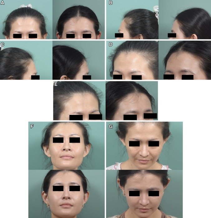
Background: Frontal fullness in Asians is often considered to indicate one's public popularity and leadership skills. Numerous materials and techniques have been applied clinically to recontour or volumize the frontal area, with variable results. The micro-autologous fat transplantation (MAFT) technique proposed by Lin et al. (2nd academic congress of Taiwan Cosmetic Association Taipei, Taiwan) in 2007 has demonstrated its feasibility in facial rejuvenation. In the present study, we used an innovative instrument to apply the MAFT technique to frontal augmentation with fat grafting and reported the results.

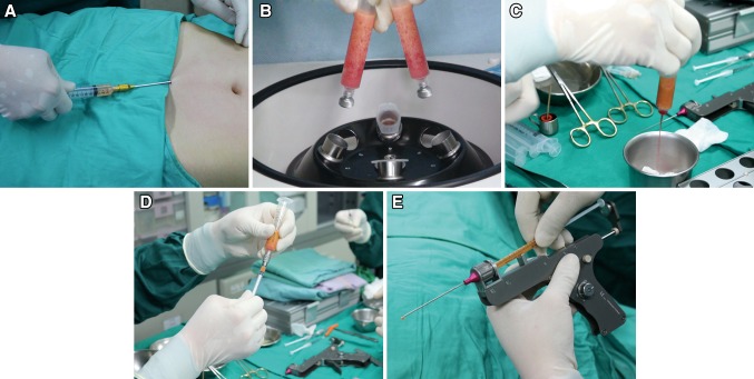
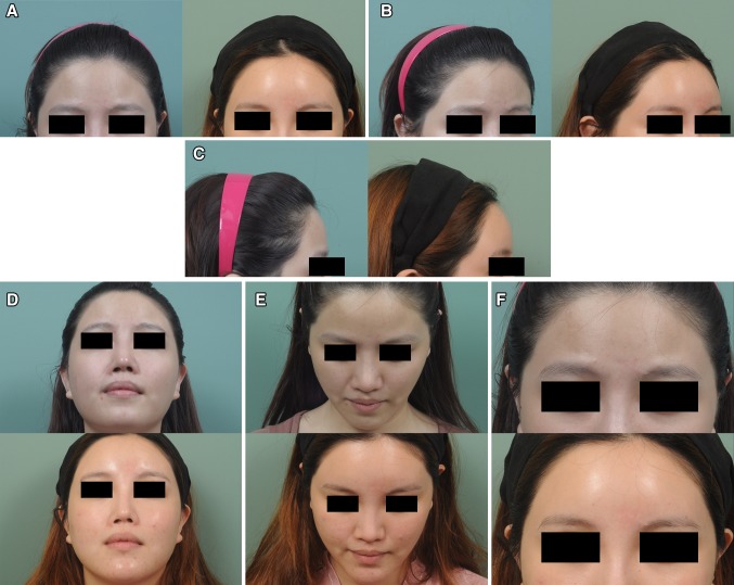
Methods: MAFT was performed on 178 patients (167 female, 11 male) during a 5-year period starting in January 2010. Fat was harvested by liposuction, processed and refined by centrifugation at 1200×g for 3 min. The purified fat was micro-transplanted for frontal contouring with the assistance of an instrument, the MAFT-GUN. The patients were followed up regularly, and photographs were taken for comparison.

Results: On average, the MAFT procedure took 52 min to complete. The average amount of delivered fat was 10.2 mL. The follow-up period was 34 months on average. No complications, including neurovascular injury, skin necrosis, abscess, nodulation, calcification or irregularity, were noted. A patient-rated satisfaction 5-point Likert scale demonstrated that 83.1% of all patients had favorable results (48.3% were satisfied, and 34.8% were very satisfied).

Conclusion: The concept and technique of MAFT has changed fat grafting from an operation with unpredictable clinical results to an easy, reliable and consistent procedure. Furthermore, the use of a precisely controlled instrument enabled surgeons to perform highly accurate micro-fat grafting. In comparison with other strategies for volume restoration, the MAFT procedure demonstrated high patient satisfaction with the long-term results. Therefore, the use of MAFT as an alternative approach to forehead contouring and volumizing was addressed.

Level of evidence iv: This journal requires that authors assign a level of evidence to each article. For a full description of these Evidence-Based Medicine ratings, please refer to the Table of Contents or the online Instructions to Authors www.springer.com/00266 .

Keywords: Fat graft; Forehead; Micro-autologous fat transplantation (MAFT).


Conflict of interest statement

Dr. Tsai-Ming Lin owns the patent rights of the MAFT-GUN and is the scientific adviser of Dermato Plastica Beauty Co., the manufacturer of the MAFT-GUN device. None of the other authors have any financial disclosures or conflicts of interest.

Figures

Fig. 1
Fig. 1
a The surgical planning of forehead volumizing and contouring is outlined as a brown shadow. The X point is the insertion site made using a #11 blade. b The deep layer is highlighted in blue (supra-periosteum, a space between the frontal periosteum and frontalis muscle) to be bounded by the frontal hairline, bilateral temporal ridges and super-orbital ridges. c The middle layer in green (intra-frontalis muscle) is located between the frontalis muscles of the forehead. d The superficial subcutaneous layer is shown in pink and is designed to act as a smoothing agent over the forehead boundary
Fig. 2
Fig. 2
a Fat aspiration is performed by back-pulling the plunger of a 10-mL syringe to approximately 2–3 mL to maintain negative pressure. b The centrifugation is maintained at 3000 rpm (approximately 1200×g) for 3 min to process and purify the lipoaspirate. c The lower part of the centrifuged lipoaspirate (bloody content) is leaked out, and the oil is wiped off using gauze on the upper part. d The purified, condensed fat is transferred to a 1-mL Luer-slip syringe for transplantation. e The fat-filled syringe is loaded into the MAFT-GUN for the MAFT procedure. The six-graded dial is set at 120, indicating that the delivered volume of each fat parcel is 1/120 mL (0.0083 mL)
Fig. 3
Fig. 3
The coronal section of the forehead is shown to illustrate the deeper layer (blue), middle layer (green) and superficial layer (pink) with the transplantation of micro-fat parcels with a size of 1/120 mL (0.0083 mL)
Fig. 4
Fig. 4
(Case 1) This 54-year-old woman presented for recontouring with fat grafting to increase the youthful appearance of her forehead. MAFT was performed to place a 12-mL fat graft (Pre-op in a, b, c, d and e, left). Six months after a single MAFT session, the volume restoration of the frontal area was maintained with fullness (Post-op in a, b, c, d and e, right). Chin-up, chin-down and close-up views showed the improved contouring (Pre-op in f, g, h and i upper; Post-op, lower)
Fig. 5
Fig. 5
(Case 2) This 25-year-old woman presented for augmentation of her forehead and temporal areas with fat grafting. MAFT was performed on her frontal area to place a 20-mL fat graft (Pre-op in a, b and c, left). Two years after a single MAFT session, the volume was maintained on the frontal area (Post-op in a, b and c, right). Chin-up, chin-down and close-up views show the improved contouring of the frontal area, which shows a smooth and abundant appearance (Pre-op in d, e and f upper; Post-op, lower)
Fig. 6
Fig. 6
(Case 3) This 33-year-old woman presented for fat grafting to restore her frontal contour. MAFT was performed to place a 10-mL fat graft (Pre-op in a, b and c, left). Six months after a single MAFT session, the fullness and volume restoration of the hollowing frontal area were maintained (Post-op in a, b and c, right). The improved appearance of frontal unevenness and the rejuvenating effect of the skin are illustrated in a close-up quarter view (Pre-op in d and e, left; Post-op in right). Chin-up, chin-down and close-up views showed the recontouring of her uneven frontal area (Pre-op in f, g and h, upper; Post-op in lower)
Fig. 7
Fig. 7
The evolution of the parcel size (volume) was initiated in 1893 by Neuber. The use of the 1/10 mL Rachet Gun per fat parcel (even l mL by larger Rachet Gun) was popular in the 1970–1980s. In 1993, Dr. Coleman proposed that the injection volume for each parcel should be 1/50th–1/30th mL. Moreover, in 1993–1994, Dr. Carpaneda’s theory proposed only a 40% survival rate in the peripheral zone 1.5 ± 0.5 mm of the parcel margin. The conceptualization of MAFT (micro-autologous fat transplantation) was advocated by Dr. Lin et al. in 2006. MAFT emphasizes that each delivered fat parcel is ideally smaller than 1/100 mL (0.01 mL), rendering the real radius of such a parcel to be 1.3 mm
Fig. 8
Fig. 8
a Three dye colors are injected into different layers (blue in deep, green in middle and pink in superficial layer) of the forehead area, as shown in the cadaver dissection. b Close-up view demonstrating that the blue dye is in the deep supra-periosteum layer, the green dye is shown in the middle intra-muscular layer, and the pink dye is distributed in the superficial subcutaneous layer

Comment in

Similar articles

Cited by

References

    1. Knize David M. The forehead and temporal fossa; anatomy and technique. Philadelphia: Lippincott Williams & Wilkins; 2011.
    1. Sykes JM. Applied anatomy of the temporal region and forehead for injectable fillers. J Drugs Dermatol. 2009;8(10 Suppl):s24–s27. - PubMed
    1. Isik S, Sahin I. Contour restoration of the forehead by lipofilling: our experience. Aesthet Plast Surg. 2012;36(4):761–766. doi: 10.1007/s00266-011-9800-2. - DOI - PubMed
    1. Hönig JF, Merten HA, Nitsch A, Verheggen R. Contouring of cranial vault irregularities with hydroxyapatite cement: a clinical and experimental investigation. J Craniofac Surg. 2005;16:457–460. doi: 10.1097/01.SCS.0000147392.91369.71. - DOI - PubMed
    1. Yaremchuk MJ. Facial skeletal reconstruction using porous polyethylene implants. Plast Reconstr Surg. 2003;111:1818–1827. doi: 10.1097/01.PRS.0000056866.80665.7A. - DOI - PubMed