. 2017 Aug;44(8):1393-1407.
doi: 10.1007/s00259-017-3683-7. Epub 2017 Apr 27.

Accuracy of diagnostic imaging modalities for peripheral post-traumatic osteomyelitis - a systematic review of the recent literature

Affiliations

Accuracy of diagnostic imaging modalities for peripheral post-traumatic osteomyelitis - a systematic review of the recent literature

Geertje A Govaert et al. Eur J Nucl Med Mol Imaging. 2017 Aug.

Abstract

Aims: Post-traumatic osteomyelitis (PTO) is difficult to diagnose and there is no consensus on the best imaging strategy. The aim of this study is to present a systematic review of the recent literature on diagnostic imaging of PTO.

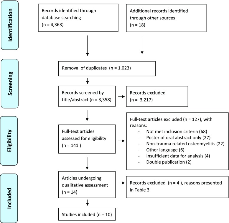
Methods: A literature search of the EMBASE and PubMed databases of the last 16 years (2000-2016) was performed. Studies that evaluated the accuracy of magnetic resonance imaging (MRI), three-phase bone scintigraphy (TPBS), white blood cell (WBC) or antigranulocyte antibody (AGA) scintigraphy, fluorodeoxyglucose positron emission tomography (FDG-PET) and plain computed tomography (CT) in diagnosing PTO were considered for inclusion. The review was conducted using the PRISMA statement and QUADAS-2 criteria.

Results: The literature search identified 3358 original records, of which 10 articles could be included in this review. Four of these studies had a comparative design which made it possible to report the results of, in total, 17 patient series. WBC (or AGA) scintigraphy and FDG-PET exhibit good accuracy for diagnosing PTO (sensitivity ranged from 50-100%, specificity ranged from 40-97% versus 83-100% and 51%-100%, respectively). The accuracy of both modalities improved when a hybrid imaging technique (SPECT/CT & FDG-PET/CT) was performed. For FDG-PET/CT, sensitivity ranged between 86 and 94% and specificity between 76 and 100%. For WBC scintigraphy + SPECT/CT, this is 100% and 89-97%, respectively.

Conclusions: Based on the best available evidence of the last 16 years, both WBC (or AGA) scintigraphy combined with SPECT/CT or FDG-PET combined with CT have the best diagnostic accuracy for diagnosing peripheral PTO.

Keywords: Antigranulocyte antibody scintigraphy; CT scan; Diagnostic imaging; FDG-PET; Fracture; Fracture related infection; MRI; Open reduction and internal fixation (ORIF); Osteosynthetic material; Ostheosynthesis; Post-traumatic osteomyelitis; White blood cell scintigraphy.


Conflict of interest statement

Ethical approval

This article does not contain any studies with human participants performed by any of the authors.

Conflict of interest

None

Figures

Fig. 1
Fig. 1
PRISMA Flow diagram
Fig. 2
Fig. 2
Clinical example of WBC scintigraphy + SPECT/CT. A 37-year-old man with a grade 3A complicated distal humeral fracture of the left elbow, initially treated with an external fixator and subsequently by plate osteosynthesis of the distal humerus. c X-ray: situation after recent fixation of the fracture with plate osteosynthesis, no signs of loosening or infection. After 4 months, he presented with a fistula and a clinical suspicion of osteomyelitis of the distal humerus. a–b, d–e: WBC scintigraphy (a image at 4 hours, b image at 24 hours, d–e fusion SPECT/CT images) after the injection of 220 MBq 99 m-Tc-labeled leucocytes demonstrated an infection around the implant at the lateral side of the elbow/distal screw. The low uptake points to only a low-grade appearance and the location to soft tissue involvement; this was confirmed at operation (f clinical pre-operative picture, g perioperative clinical picture)
Fig. 3
Fig. 3
Clinical example of FDG-PET/CT. A 77-year-old woman who had a proximal femur fracture for which she underwent open reduction and internal fixation with a femur plate which had to be removed at a later stage due to infection. a X-ray, AP view: no consolidation, severe angulation, heterogeneous sclerotic aspect around the fracture. She was referred to our hospital with a fistula in the lateral thigh and a clinical suspicion of osteomyelitis of the proximal femur. Further imaging demonstrated an infection of the proximal femur, a medial abscess and a fistula coursing to the lateral aspect of the thigh which correlated with the clinical findings during surgery. b–f 18F FDG-PET/CT (b coronal FDG-PET image, c coronal fused FDG-PET/CT image, d–f transaxial fused FDG-PET/CT images). g clinical pre-operative picture, h perioperative clinical picture
Fig. 4
Fig. 4
Clinical example of MRI. A 54-year-old man with a history of an open fracture treated with a plate many years ago. The fracture healed slowly and then the plate was removed because of continued skin breakdown over the front of the tibia. a Frontal and lateral radiograph demonstrating sclerosis and chronic periosteal reaction around the previous fracture site. b Sagittal fat-suppressed images of the calf demonstrating bone and soft tissue oedema. c & d Axial fat-suppressed images demonstrating sequestra (blue arrow), cortical abscesses (yellow arrows) and periostitis and soft tissue oedema (red arrow)

References

    1. McNally M, Sendi P. Implant associated osteomyelitis of the long bones. In: Zimmerli W, editor. Bone and Joint Infections: from microbiology to diagnostics and treatment.: Wiley-Blackwell; 2015. p. 303–23.
    1. Trampuz A, Zimmerli W. Diagnosis and treatment of infections associated with fracture-fixation devices. Injury. 2006;37(Suppl 2):S59–66. doi: 10.1016/j.injury.2006.04.010. - DOI - PubMed
    1. Bose D, Kugan R, Stubbs D, McNally M. Management of infected nonunion of the long bones by a multidisciplinary team. The bone & joint journal. 2015;97-B(6):814–7. doi: 10.1302/0301-620X.97B6.33276. - DOI - PubMed
    1. Korim MT, Payne R, Bhatia M. A case-control study of surgical site infection following operative fixation of fractures of the ankle in a large U.K. trauma unit. The bone & joint journal. 2014;96-B(5):636–40. doi: 10.1302/0301-620X.96B5.33143. - DOI - PubMed
    1. Ktistakis I, Giannoudi M, Giannoudis PV. Infection rates after open tibial fractures: are they decreasing? Injury. 2014;45(7):1025–7. doi: 10.1016/j.injury.2014.03.022. - DOI - PubMed

Publication types

MeSH terms