Review
. 2017 Jul;52(7):767-776.
doi: 10.1007/s00535-017-1342-1. Epub 2017 Apr 27.

Reflux esophagitis and its role in the pathogenesis of Barrett's metaplasia

Affiliations
Review

Reflux esophagitis and its role in the pathogenesis of Barrett's metaplasia

Rhonda Frances Souza. J Gastroenterol. 2017 Jul.

Abstract

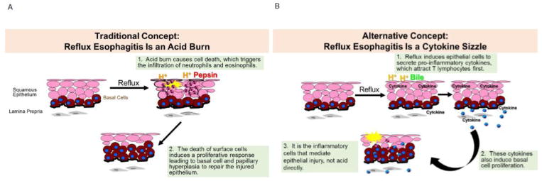
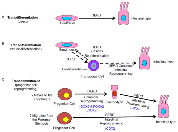
Reflux esophagitis damages the squamous epithelium that normally lines the esophagus, and promotes replacement of the damaged squamous lining by the intestinal metaplasia of Barrett's esophagus, the precursor of esophageal adenocarcinoma. Therefore, to prevent the development of Barrett's metaplasia and esophageal adenocarcinoma, the pathogenesis of reflux esophagitis must be understood. We have reported that reflux esophagitis, both in a rat model and in humans, develops as a cytokine-mediated inflammatory injury (i.e., cytokine sizzle), not as a caustic chemical injury (i.e., acid burn), as traditionally has been assumed. Moreover, reflux induces activation of hypoxia inducible factor (HIF)-2α, which enhances the transcriptional activity of nuclear factor kappa-light-chain-enhancer of activated B cells (NF-κB) causing increases in pro-inflammatory cytokines and in migration of T lymphocytes, an underlying molecular mechanism for this cytokine-mediated injury. In some individuals, reflux esophagitis heals with Barrett's metaplasia. A number of possibilities exist for the origin of the progenitor cells that give rise to this intestinal metaplasia including those of the esophagus, the proximal stomach, or the bone marrow. However, intestinal cells are not normally found in the esophagus, the stomach, or the bone marrow. Thus, the development of Barrett's intestinal metaplasia must involve some molecular reprogramming of key developmental transcription factors within the progenitor cell, a process termed transcommitment, which may be initiated by the noxious components of the gastric refluxate. This review will highlight recent studies on the pathogenesis of reflux esophagitis and on reflux-related molecular reprogramming of esophageal squamous epithelial cells in the pathogenesis of Barrett's metaplasia.

Keywords: Barrett’s esophagus; Cdx2; Cytokine; NF-κB; Squamous cells.


Conflict of interest statement

Compliance with ethical standards

Conflict of interest The author declares that she has no conflict of interest.

Figures

Figure 1
Figure 1
Concepts on the Pathogenesis of Reflux Esophagitis. (A) The traditional concept has been that reflux esophagitis results from a caustic (acid) burn. When esophageal squamous epithelium is exposed to reflux, acid and pepsin are thought to damage the junctions between the cells, making the epithelium permeable and allowing acid to seep into the epithelium and injure the epithelial cells. This acid burn causes cell death, which triggers the infiltration of neutrophils and eosinophils into the epithelium. The death of surface cells is also assumed to induce a proliferative response leading to basal cell and papillary hyperplasia to repair the injured epithelium. (B) The alternative concept that we propose is that reflux esophagitis develops as a cytokine-mediated inflammatory injury (i.e. cytokine sizzle). In this model, the reflux of acid and bile salts doesn’t destroy epithelial cells directly, but rather induces them to secrete pro-inflammatory cytokines, which attract T lymphocytes first. These cytokines also induce basal cell proliferation. Ultimately, it is inflammatory cells that mediate the epithelial injury, not the direct acid burn.
Figure 2
Figure 2
Confocal Laser Endomicroscopy (CLE) Images Of Acute Reflux Esophagitis. Representative images from the distal esophagus of an individual study subject at baseline on proton pump inhibitors (PPIs), week 1 and week 2 off of PPIs. White arrows indicate fluorescein within the intraepithelial capillaries at baseline on PPIs. By 1 and 2 weeks off PPIs, the fluorescein has leaked from the blood vessels into the intercellular spaces enhancing the identification of the individual squamous cells. Images courtesy of Dr. Kerry B. Dunbar
Figure 3
Figure 3
Conceptual Overview of GERD-Induced Cellular Reprogramming in the Pathogenesis of Barrett’s Metaplasia. Potential pathways for the origin of Barrett’s metaplasia include (A) Direct transdifferentiation is the process in which an individual, fully differentiated cell (i.e. squamous) change directly into another type of fully differentiated cell (i.e. intestinal-type cell) in the setting of GERD. (B) Transdifferentiation in the setting of GERD may result in de-differentiated cells with features of both squamous and intestinal cell types (transitional cells). If GERD subsides, this transitional cell can re-differentiate into a squamous cell. If GERD continues, this transitional cell can reprogram into the new intestinal-type cell through a series of intervening cell divisions. (C) Transcommitment is the process in which immature progenitor cells are reprogrammed in the setting of GERD to give rise to the gastric and intestinal cell types that comprise Barrett’s metaplasia. Progenitor cells that are native to the esophagus undergo reprogramming to columnar gastric-type cells. Some of these gastric-type columnar cells undergo further reprogramming into intestinal-type cells. Progenitor cells may migrate from the proximal stomach into the esophagus, but some of these gastric progenitor cells would still have to undergo reprogramming into intestinal-type cells. Some of the transcription factors that have been implicated in these GERD-induced reprogramming processes are indicated in blue.

Similar articles

Cited by

References

    1. Spechler SJ. Carcinogenesis at the gastroesophageal junction: Free radicals at the frontier. Gastroenterology. 2002;122:1518–1520. - PubMed
    1. Spechler SJ, Souza RF. Barrett’s esophagus. The New England journal of medicine. 2014;371:836–845. - PubMed
    1. Iwakiri K, Kinoshita Y, Habu Y, Oshima T, Manabe N, Fujiwara Y, Nagahara A, Kawamura O, Iwakiri R, Ozawa S, Ashida K, Ohara S, Kashiwagi H, Adachi K, Higuchi K, Miwa H, Fujimoto K, Kusano M, Hoshihara Y, Kawano T, Haruma K, Hongo M, Sugano K, Watanabe M, Shimosegawa T. Evidence-based clinical practice guidelines for gastroesophageal reflux disease 2015. Journal of gastroenterology. 2016;51:751–767. - PubMed
    1. Spechler SJ. Clinical practice. Barrett’s esophagus The New England journal of medicine. 2002;346:836–842. - PubMed
    1. Ismail-Beigi F, Horton PF, Pope CE., 2nd Histological consequences of gastroesophageal reflux in man. Gastroenterology. 1970;58:163–174. - PubMed

MeSH terms

Substances

LinkOut - more resources