. 2017 Jul;105(2):273-293.
doi: 10.1111/mmi.13700. Epub 2017 May 18.

Use of chimeric type IV secretion systems to define contributions of outer membrane subassemblies for contact-dependent translocation

Affiliations

Use of chimeric type IV secretion systems to define contributions of outer membrane subassemblies for contact-dependent translocation

Jay E Gordon et al. Mol Microbiol. 2017 Jul.

Abstract

Recent studies have shown that conjugation systems of Gram-negative bacteria are composed of distinct inner and outer membrane core complexes (IMCs and OMCCs, respectively). Here, we characterized the OMCC by focusing first on a cap domain that forms a channel across the outer membrane. Strikingly, the OMCC caps of the Escherichia coli pKM101 Tra and Agrobacterium tumefaciens VirB/VirD4 systems are completely dispensable for substrate transfer, but required for formation of conjugative pili. The pKM101 OMCC cap and extended pilus also are dispensable for activation of a Pseudomonas aeruginosa type VI secretion system (T6SS). Chimeric conjugation systems composed of the IMCpKM101 joined to OMCCs from the A. tumefaciens VirB/VirD4, E. coli R388 Trw, and Bordetella pertussis Ptl systems support conjugative DNA transfer in E. coli and trigger P. aeruginosa T6SS killing, but not pilus production. The A. tumefaciens VirB/VirD4 OMCC, solved by transmission electron microscopy, adopts a cage structure similar to the pKM101 OMCC. The findings establish that OMCCs are highly structurally and functionally conserved - but also intrinsically conformationally flexible - scaffolds for translocation channels. Furthermore, the OMCC cap and a pilus tip protein coregulate pilus extension but are not required for channel assembly or function.


Figures

FIG. 1
FIG. 1
E. coli pKM101 tra gene deletion and complementation analyses. The E. coli pKM101 tra and A. tumefaciens virB loci are similar in gene composition and order, as shown by color-coding of genes encoding homologs of the T4SS subunits. The pKM101 tra genes expressed from pRP100 encode a fully functional Tra T4SS, as shown by efficient conjugative DNA transfer and IKe bacteriophage sensitivity. The schematic depicts effects of individual Δtra mutations (histogram, upper bars) and results of complementation studies (histogram, lower bars) in which corresponding genes were trans-expressed from the PBAD promoter (black arrow) on conjugative transfer. Matings (2 h) were carried out on solid-surface (solid bars) and in liquid (stippled bars); pRP100-carrying donors also were mated overnight in liquid with constant agitation (light stippled bars). *, denotes transfer frequencies below the threshold of detection (<10−8 Tc’s/D). Transfer frequencies are presented as transconjugants/donor (Tc’s/D). IKe phage sensitivity (S, sensitive, R, resistant) for Δtra mutants and complemented strains is shown at the right.
FIG. 2
FIG. 2
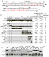
Substitution and deletion mutational analysis of the outer membrane cap of the pKM101 Tra T4SS. A) Ribbon diagram of the O-layer of the pKM101 outer membrane core complex (OMCC). VirB7-like TraN and VirB9-like TraO are color-coded magenta and cyan, respectively. The α-helical antennae projection (AP) forming the OM-spanning cap and the C-terminal (CT) domain of TraF are color-coded red, and the β-barrel domain of TraF is color-coded yellow. At right, ribbon diagram of a TraF monomer depicting the β-barrel, AP, and CT domains in same color-coding. Domain junctions (residues from N terminus) and positions of deletion or substitution mutations are indicated. B) Schematic depicts TraF domain architecture with junctions (in residues) indicated. Mutations in the AP or CT are listed at left, and effects of the mutations on plasmid transfer (transconjugants per donor, Tc’s/D) and IKe phage infection (S, sensitive; R, resistant). C) Levels of His-TraF and mutant proteins in total cell extracts, as monitored by immunostaining with α-His antibodies. RNA polymerase β-subunit (α-RNAP) served as a loading control.
FIG. 3
FIG. 3
Domain swapping reveals compositional flexibility of TraF’s β-barrel, antennae projection (AP,) and C terminus (CT). A) Sequence alignment of the AP and C-terminal (CT) domains of TraF and VirB10, with identical (red) and nonidentical (black) residues shown. Numbers correspond to domain junctions (residues from N terminus). Sequences comprising the α2 - loop (APL) - α3 regions of AP domains and the highly-conserved RDLF motifs are highlighted. B) Schematics depicting domains of TraF and VirB10, with junctions (residues from N terminus) indicated: Cyto, cytoplasmic; TM, transmembrane domain; Pro-Rich, proline-rich-region; β-Barrel; AP, antennae projection; CT, C-terminal domain. Schematics of the TraF/VirB10 chimeras depict the VirB10 domain(s) swapped for the equivalent domain(s) of TraF. Strains producing the TraF/VirB10 chimeras supported plasmid transfer in 2 h solid-surface matings at the frequencies shown in transconjugants per donor (Tc’s/D), and exhibited sensitivity (S) or resistance (R) to IKe infection. C) Levels of His-TraF and chimeric proteins in total cell extracts, as monitored by immunostaining with α-His antibodies. RNA polymerase β-subunit (α-RNAP) served as a loading control.
FIG. 4
FIG. 4
Chimeric T4SSs support conjugative DNA transfer and activate T6SS killing. A) Sequences encoding the outer membrane core complex (OMCC) subunits TraN, TraO, and the C-terminal half (residues 194–386) of TraF were replaced with corresponding genes or gene fragments from the A. tumefaciens VirB, E. coli R388 Trw, or B. pertussis Ptl systems on mini-pKM101 plasmid pCGR108. The chimeric T4SSs composed of the inner membrane complex (IMC) of pKM101 (yellow) joined to the OMCCs from the VirB, Trw, or Ptl systems (color-coded) are modeled on the R388 T4SS3–10 structure (Low et al., 2014). B) E. coli donors carrying pCGR108 derivatives encoding the only the TrapKM101 IMC or OMCC, or the IMC::OMCC chimeras transferred the mobilizable plasmid pJG42 at the frequencies shown in transconjugants per donor (Tc’s/D) in solid-surface (histogram, solid bars) or liquid (stippled bars) matings, and were resistant to IKe phage infection (S, sensitive; R, resistant). C) E. coli survival when cultivated in the absence or presence of P. aeruginosa PAO1. E. coli DH5α cells lacked or produced intact or variant forms of the TrapKM101 T4SS depicted. Statistical significance is shown based on a Student’s t test corresponding to the values of plasmid-free DH5α or growth in the absence of P. aeruginosa (NS, not significant; *P < 0.05; **P < 0.01). For panels B and C, data presented are mean +/− SD, n = 3 independent replicates.
FIG. 5
FIG. 5
Requirements for activation of T6SS killing by P. aeruginosa PAO1. E. coli DH5α lacking or producing the TrapKM101 T4SS composed of the His6-TraF variants shown; in each case, the traF allele was substituted for wild-type traF by incorporation into the pKM101 tra locus on plasmid pCGR108. Statistical significance is shown based on a Student’s t test corresponding to the values of plasmid-free DH5α or growth in the absence of P. aeruginosa (NS, not significant; *P < 0.05; **P < 0.01). Data presented are mean +/− SD, n = 3 independent replicates. Lower panel: Levels of His-TraF variants in total cell extracts, as monitored by immunostaining with α-His antibodies. RNA polymerase β-subunit (α-RNAP) served as a loading control.
FIG. 6
FIG. 6
Negative-stain EM structure of the A. tumefaciens outer-membrane core complex (OMCC) and comparison with the NS-EM structure of the OMCC (EMDB-5032) encoded by E.coli pKM101. A) A. tumefaciens OMCC side view (left) and cut-away side view (right). B) E.coli pKM101 OMCC side view (left) and cut-away side view (right). C) Representation of the cut-away side view of the overlay of A. tumefaciens and E.coli pKM101 OMCC’s. D) Cross-section of overlaid A. tumefaciens and E.coli pKM101 OMCC complexes. Dashed line S in panel C indicates the level of the cross section shown in panel D.
FIG. 7
FIG. 7
Working model for biogenesis of Type IVa secretion systems highlighting the importance of the postulated OMCC checkpoint in regulating pilus extension. Steps in the assembly pathway of the T4SS include (A) formation of the stable T4SS3–10 substructure (Low et al., 2014) and (B) elaboration of a short pilus that extends from an inner membrane platform to the cell surface by a mechanism requiring TraB/VirB4- and TraG/VirB11-type ATPases. Next, (C) the pilus extends from the cell surface in a mate-seeking mode by a mechanism activated by recruitment of surface-exposed TraC to the distal end of the OMCC (denoted by yellow lightning bolt). TraC alternatively might be recruited to the T4SS via a periplasmic location (red-dashed line, ?). Finally, (D) upon pilus-mediated or direct contact with a recipient cell, a mating signal is transduced across the donor cell envelope resulting in recruitment of the TraJ/VirD4 substrate receptor, substrate docking and ATPase hydrolysis. These signals (denoted by lightning bolts) activate the morphogenetic switch to the T4SS ‘mating’ mode. The assembly intermediate depicted in (B) may bypass the pilus assembly (mate-seeking) mode (C) if presented with signals, e.g., recipient cell contact, required for activation of the substrate transfer (mating) mode (D), as could occur when donors and recipients grow in dense biofilm (solid-surface) communities. Abbreviations: OM, outer membrane; IM, inner membrane; P, peptidoglycan; OMCC, outer membrane complex; IMC, inner membrane complex; GSP, general secretory pathway. The pKM101 Tra proteins and their VirB counterparts required for each step of the assembly pathway are denoted.

Similar articles

Cited by

References

    1. Aly KA, Baron C. The VirB5 protein localizes to the T-pilus tips in Agrobacterium tumefaciens. Microbiology. 2007;153:3766–3775. - PubMed
    1. Ames BN, McCann J, Yamasaki E. Methods for detecting carcinogens and mutagens with the Salmonella/mammalian-microsome mutagenicity test. Mutat Res. 1975;31:347–364. - PubMed
    1. Arutyunov D, Frost LS. F conjugation: back to the beginning. Plasmid. 2013;70:18–32. - PubMed
    1. Atmakuri K, Cascales E, Christie PJ. Energetic components VirD4, VirB11 and VirB4 mediate early DNA transfer reactions required for bacterial type IV secretion. Mol Microbiol. 2004;54:1199–1211. - PMC - PubMed
    1. Babic A, Lindner AB, Vulic M, Stewart EJ, Radman M. Direct visualization of horizontal gene transfer. Science. 2008;319:1533–1536. - PubMed

MeSH terms