

Evaluation of the osseointegration of dental implants coated with calcium carbonate: an animal study
- PMID: 28452375
- PMCID: PMC5709541
- DOI: 10.1038/ijos.2017.13
Evaluation of the osseointegration of dental implants coated with calcium carbonate: an animal study
Abstract
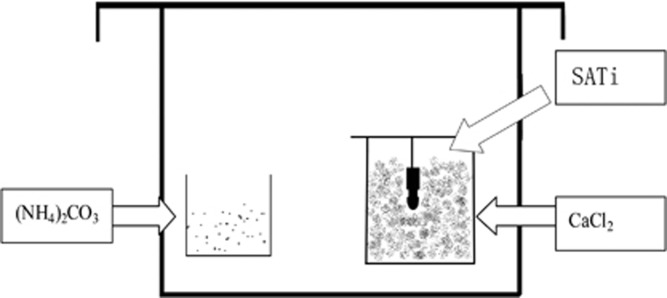
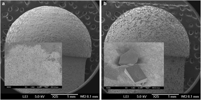
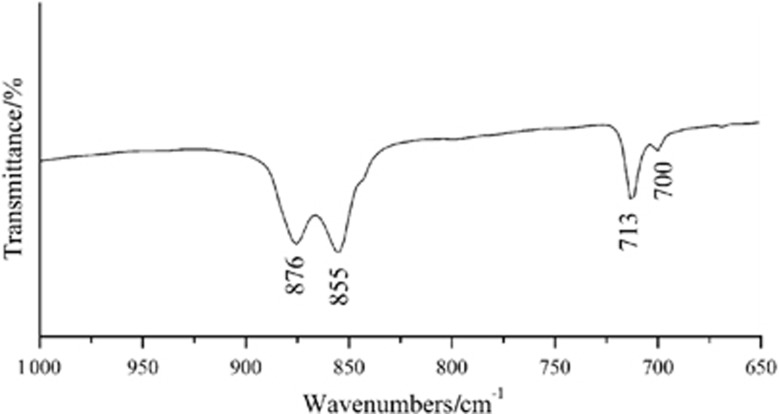
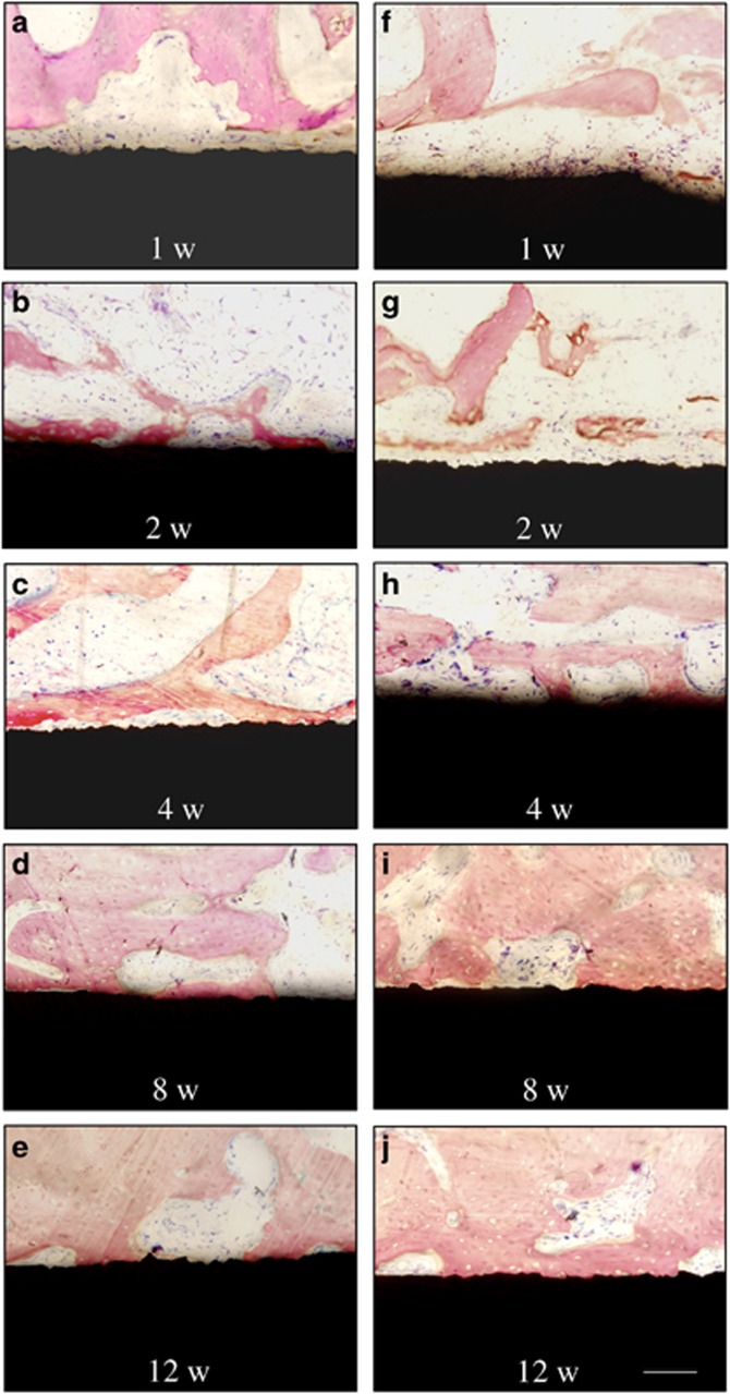
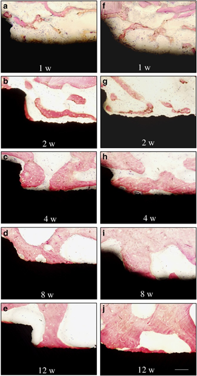
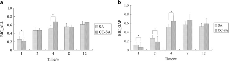
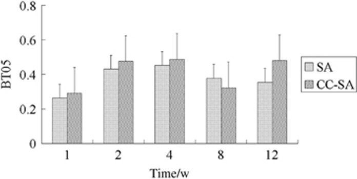
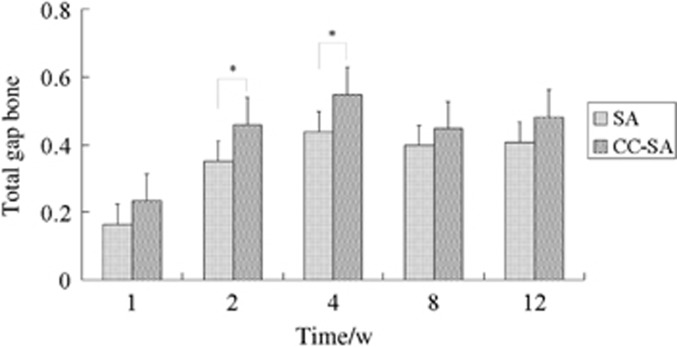
In an attempt to overcome the limitations of titanium in dental and orthopaedic clinical applications, a new method has been developed to prepare calcium carbonate coatings on sandblasted and acid-etched (SA) titanium implants. The purpose of this study was to investigate the effect of calcium carbonate-SA (CC-SA) implants on osseointegration in vivo. The surfaces of SA and CC-SA implants were characterised for surface morphology and surface chemistry. Subsequently, these two kinds of implants were implanted in the femoral condyles of rabbits. The implants were retrieved and prepared for histological and histomorphometric evaluation 1, 2, 4, 8 and 12 weeks after implantation. Significantly higher values of bone-to-implant contact of the entire implant except the gap area (BIC_ALL) and the bone-to-implant contact of the gap area (BIC_GAP) were found in animals with the CC-SA implants than in those with the SA implants at 4 weeks. Higher values of total gap bone were found in those with the CC-SA implants than in those with the SA implants at 1, 2 and 4 weeks. In conclusion, the current findings demonstrate that the calcium carbonate coating can improve and accelerate the early ingrowth of bone and osseointegration at the early healing phase. This may reduce clinical healing times and thus improve implant success rates.
Figures
References
-
- Nebe JB, Mueller L, Luethen F et al. Osteoblast response to biomimetically altered titanium surfaces. Acta Biomater 2008; 4 (6): 1985–1995. - PubMed
-
- Bacchelli B, Giavaresi G, Franchi MA et al. Influence of a zirconia sandblasting treated surface on peri-implant bone healing: an experimental study in sheep. Acta Biomater 2009; 5 (6): 2246–2257. - PubMed
-
- Park JW, Suh JY, Chung HJ. Effects of calcium ion incorporation on osteoblast gene expression in MC3T3-E1 cells cultured on microstructured titanium surfaces. J Biomed Mater Res A 2008; 86 (1): 117–126. - PubMed
-
- Kokubo T, Kim HM, Kawashita M. Novel bioactive materials with different mechanical properties. Biomaterials 2003; 24 (13): 2161–2175. - PubMed
-
- Langhoff JD, Voelter K, Scharnweber D et al. Comparison of chemically and pharmaceutically modified Titanium and zirconia implant surfaces in dentistry: a study in sheep. Int J Oral Maxillofac Surg 2008; 37 (12): 1125–1132. - PubMed
Publication types
MeSH terms
Substances
LinkOut - more resources
Full Text Sources
Other Literature Sources
Miscellaneous
