Review
. 2017 Jan 2;5(1):e1274354.
doi: 10.1080/21688370.2016.1274354. Epub 2017 Jan 3.

Interactions of Giardia sp. with the intestinal barrier: Epithelium, mucus, and microbiota

Affiliations
Review

Interactions of Giardia sp. with the intestinal barrier: Epithelium, mucus, and microbiota

Thibault Allain et al. Tissue Barriers. .

Abstract

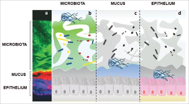
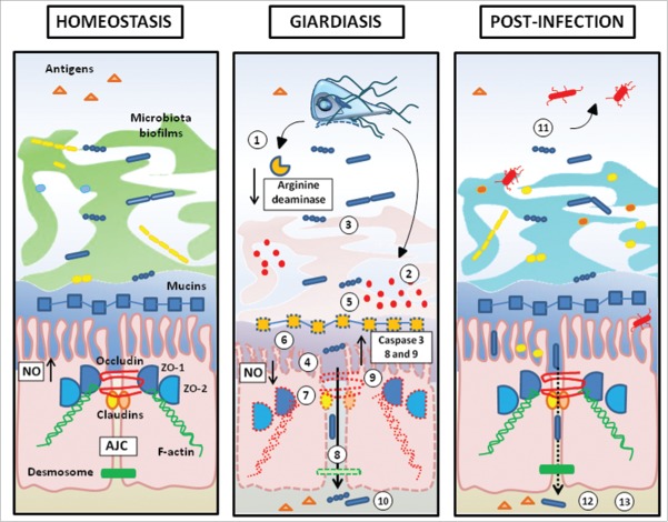
Understanding how intestinal enteropathogens cause acute and chronic alterations has direct animal and human health perspectives. Significant advances have been made on this field by studies focusing on the dynamic crosstalk between the intestinal protozoan parasite model Giardia duodenalis and the host intestinal mucosa. The concept of intestinal barrier function is of the highest importance in the context of many gastrointestinal diseases such as infectious enteritis, inflammatory bowel disease, and post-infectious gastrointestinal disorders. This crucial function relies on 3 biotic and abiotic components, first the commensal microbiota organized as a biofilm, then an overlaying mucus layer, and finally the tightly structured intestinal epithelium. Herein we review multiple strategies used by Giardia parasite to circumvent these 3 components. We will summarize what is known and discuss preliminary observations suggesting how such enteropathogen directly and/ or indirectly impairs commensal microbiota biofilm architecture, disrupts mucus layer and damages host epithelium physiology and survival.

Keywords: Giardia duodenalis; Giardiasis; commensals; host-parasite interactions; intestinal microbiota biofilm; mucus layer; poly-microbial infection.


Figures

Figure 1.
Figure 1.
Giardia interactions with gut triple barrier (microbiota, mucus, epithelium). a) characterization of intestinal barriers geographical distribution in mice immunostaining Colonic sections were stained with EUB-388 (bacteria; αDNA), WGA (mucus; Life Technologies) and DAPI (epithelial cells; SIGMA). b) commensals are mostly organized in biofilms throughout the gastrointestinal tract. It has been observed that this resident microbiota plays a role in host susceptibility to Giardia infection. Some commensals (ex: lactic acid bactaria) even exhibit anti-giardial effects. In turn, Giardia has the ability to modulate commensals to pathobionts by inducing virulence factors and also disrupting intestinal biofilms. The resulting shift of gut microbiota may help explain the production of post- infectious symptoms. c) To attach the epithelium, trophozoites must breach the mucus layer, which acts as a biochemical / physical barrier. Little is known regarding the role of mucus during Giardia infection. Ongoing research indicates that Giardia’s proteolytic activity may disrupt MUC2 mucin, the major constituent of intestinal mucus in humans; d) Trophozoites strongly attach to epithelial microvilli, and disrupt the epithelial barrier. Disaccharidase deficiencies, diffuse microvillous shortening, arginine starvation, increased permeability, disruption of tight junctions and enterocyte induced apoptosis have been associated with Giardia infection. Extracellular factors such as cathepsin B-like cysteine proteases contribute to the parasite virulence by degrading CXCL-8 (IL-8) and inducing villin breakdown.
Figure 2.
Figure 2.
Disruption of intestinal biofilm following Giardia exposure. Human microbiota biofilms from colonic biopsies were cultured ex vivo in the Calgary Biofilm Device, and exposed to vehicle (Control) or to live Giardia trophozoites (G. duodenalis). G. duodenalis depletes microbiota biofilms of their extracellular matrix coat. Representative Scanning Electron micrographs (13000 x magnification). Modified from Beatty et al. © Andre Buret. Reproduced by permission of Andre Buret. Permission to reuse must be obtained from the rightsholder.
Figure 3.
Figure 3.
Barrier disruption in giardiasis and post-infection consequences. (1) Consumption of arginine via high arginine deaminase activity. Arginine starvation leads to an impaired secretion of anti-giardial Nitric Oxide (NO); (2) Giardia’s proteolytic activity (cathepsin-B-like cysteine proteases) lead to (3) An impairment of commensal microbiota biofilms, (4) the cleavage of pro-inflamatory chemokines (CXCL-8), (5) The disruption of MUC2 mucin integrity; (6) Diffuse shortening of brush border microvilli; (7) Disruption and/or rearrangement of the apical junction complex (AJC) (ZO-1, ZO-2, claudin-1, claudin-4, occludin), filamentous actin (F-actin and α-actinin), and at desmosomal level (desmocollin); (8) Bacterial and antigen translocation in the lamina propria; (9) Induction of pro-apoptotic factors caspase-3, 8 and 9, BAX, PARP, and impairment of anti-apoptotic protein Bcl-2; (10) Immune response in giardiasis is reviewed in Einarsson et al. 2016; (11) Toxic effects of pathobionts released by dysbiotic microbiota; (12) Paracellular translocation (13) Activation of pathogenic endocrine and immunological signals.

References

    1. Halliez MC, Buret AG. Extra-intestinal and long term consequences of giardia duodenalis infections. World J Gastroenterol 2013; 19(47):8974-85; PMID:24379622; http://dx.doi.org/ 10.3748/wjg.v19.i47.8974 - DOI - PMC - PubMed
    1. Halliez MC, Motta JP, Feener TD, Guerin G, LeGoff L, Francois A, Colasse E, Favennec L, Gargala G, Lapointe TK et al.. Giardia duodenalis induces paracellular bacterial translocation and causes postinfectious visceral hypersensitivity. Am J Physiol Gastrointest Liver Physiol 2016; 310(8):G574-585; PMID:26744469. - PMC - PubMed
    1. Savioli L, Smith H, Thompson A. Giardia and cryptosporidium join the ‘neglected diseases initiative’. Trends Parasitol 2006; 22(5):203-208; PMID:16545611; http://dx.doi.org/ 10.1016/j.pt.2006.02.015 - DOI - PubMed
    1. Slavin I, Saura A, Carranza PG, Touz MC, Nores MJ, Lujan HD. Dephosphorylation of cyst wall proteins by a secreted lysosomal acid phosphatase is essential for excystation of giardia lamblia. Mol Biochem Parasitol 2002; 122(1):95-8; PMID:12076774; http://dx.doi.org/ 10.1016/S0166-6851(02)00065-8 - DOI - PubMed
    1. Ankarklev J, Jerlstrom-Hultqvist J, Ringqvist E, Troell K, Svard SG. Behind the smile: Cell biology and disease mechanisms of giardia species. Nat Rev Microbiol 2010; 8(6):413-22; PMID:20400969. - PubMed

Publication types

LinkOut - more resources