. 2017 Apr 28;18(5):926.
doi: 10.3390/ijms18050926.

Estrogen Modulates Specific Life and Death Signals Induced by LH and hCG in Human Primary Granulosa Cells In Vitro

Affiliations

Estrogen Modulates Specific Life and Death Signals Induced by LH and hCG in Human Primary Granulosa Cells In Vitro

Livio Casarini et al. Int J Mol Sci. .

Abstract

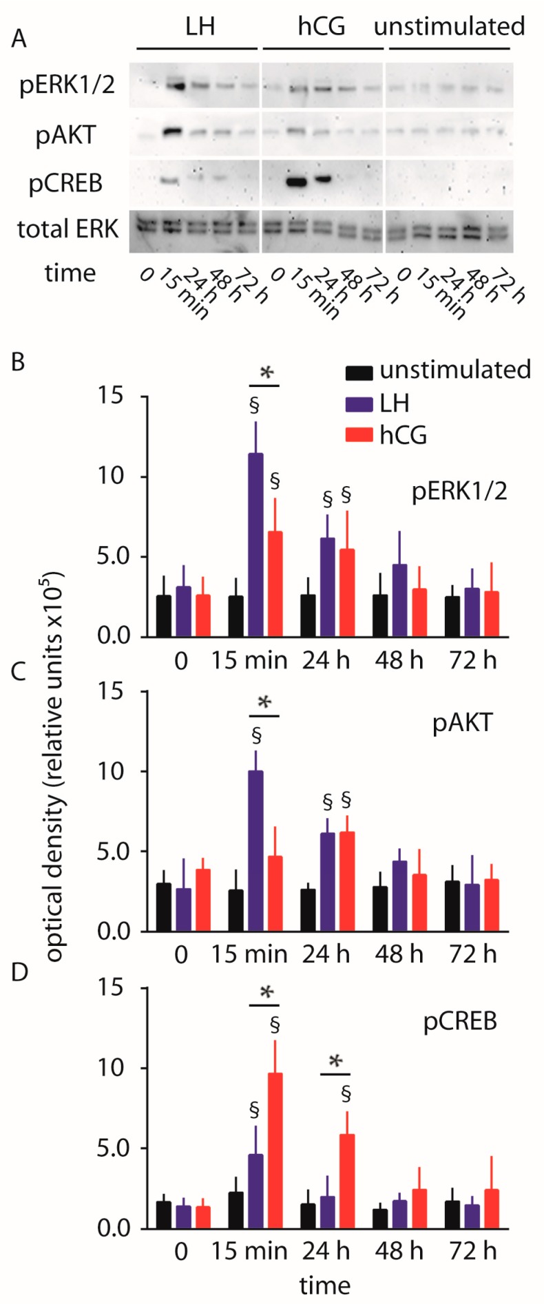
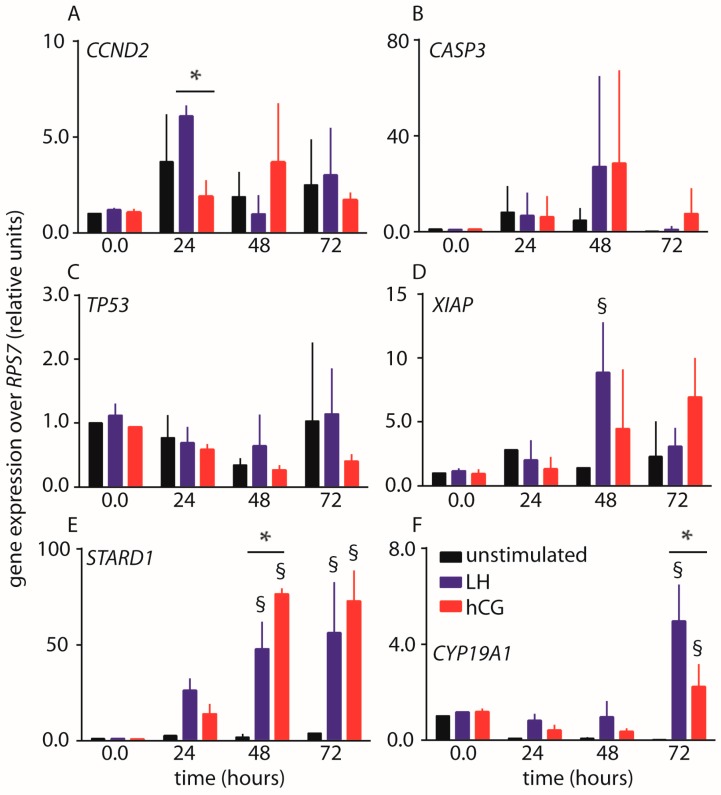
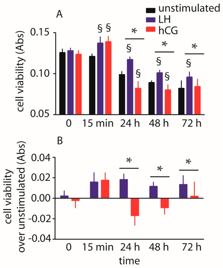
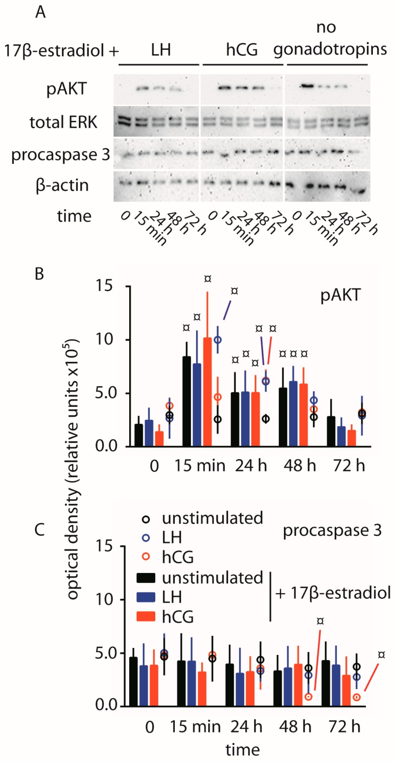
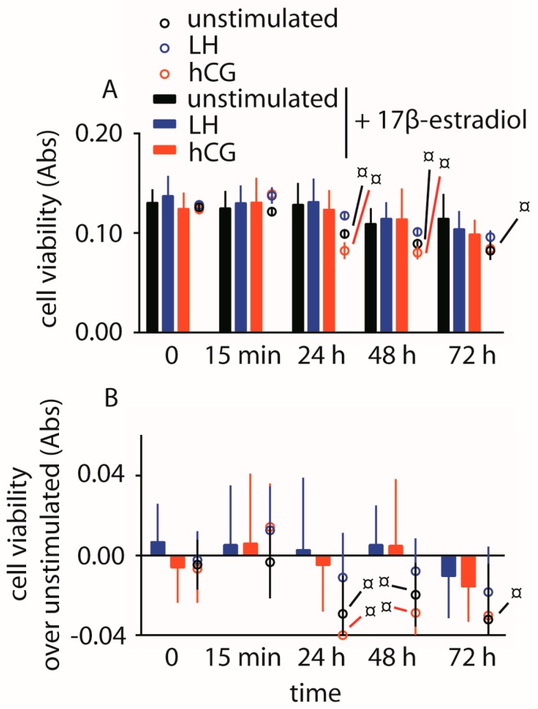
Luteinizing hormone (LH) and human chorionic gonadotropin (hCG) are glycoprotein hormones used for assisted reproduction acting on the same receptor (LHCGR) and mediating different intracellular signaling. We evaluated the pro- and anti-apoptotic effect of 100 pM LH or hCG, in the presence or in the absence of 200 pg/mL 17β-estradiol, in long-term, serum-starved human primary granulosa cells (hGLC) and a transfected granulosa cell line overexpressing LHCGR (hGL5/LHCGR). To this purpose, phospho-extracellular-regulated kinase 1/2 (pERK1/2), protein kinase B (pAKT), cAMP-responsive element binding protein (pCREB) activation and procaspase 3 cleavage were evaluated over three days by Western blotting, along with the expression of target genes by real-time PCR and cell viability by colorimetric assay. We found that LH induced predominant pERK1/2 and pAKT activation STARD1, CCND2 and anti-apoptotic XIAP gene expression, while hCG mediated more potent CREB phosphorylation, expression of CYP19A1 and procaspase 3 cleavage than LH. Cell treatment by LH is accompanied by increased (serum-starved) cell viability, while hCG decreased the number of viable cells. The hCG-specific, pro-apoptotic effect was blocked by a physiological dose of 17β-estradiol, resulting in pAKT activation, lack of procaspase 3 cleavage and increased cell viability. These results confirm that relatively high levels of steroidogenic pathway activation are linked to pro-apoptotic signals in vitro, which may be counteracted by other factors, i.e., estrogens.

Keywords: LH; apoptosis; gonadotropins; granulosa; hCG.


Conflict of interest statement

The authors declare no conflict of interest.

Figures

Figure 1
Figure 1
Comparison of luteinizing hormone (LH)- and human chorionic gonadotropin (hCG)-induced extracellular-regulated kinase 1/2 (ERK1/2), protein kinase B (AKT) and cAMP-responsive element binding protein (CREB) phosphorylation over 72 h, in human primary granulosa lutein cells (hGLC). (A) Evaluation of pERK1/2, pAKT and pCREB activation by Western blotting. Total ERK served as loading control (images representative of four independent experiments); (BD) Semi-quantification of pERK1/2, pAKT and pCREB Western blotting signals. § = significantly different to unstimulated (control) at the same time-point; * = significant difference of LH versus hCG; two-way ANOVA and Bonferroni post-test (p < 0.05; means ± standard deviation (SD); n = 4).
Figure 2
Figure 2
Evaluation of LH- and hCG-induced procaspase 3 cleavage over 72 h, in hGLC. (A) Evaluation of procaspase 3 by Western blotting. β-actin was the loading control (images representative of four independent experiments); (B) Semi-quantification of procaspase 3 Western blotting signals. § = significantly different versus unstimulated (control) samples collected at the same time-point; two-way ANOVA and Bonferroni post-test (p < 0.05; means ± SD; n = 4).
Figure 3
Figure 3
Time-course (0–72 h) expression analysis of LH- and hCG-target genes by real-time PCR. Values were normalized over the expression of RPS7 housekeeping gene. (A) CCND2 gene encoding for the cyclin D2; (B) CASP3 gene encoding for the procaspase 3; (C) TP53 gene encoding for the p53 tumor-suppressor protein; (D) XIAP gene encoding for the X-linked inhibitor of apoptosis factor; (E) STARD1 gene encoding for the StAR enzyme; (F) CYP19A1 gene encoding for the aromatase enzyme. § = significantly different to unstimulated (control) at the same time-point; * = significant difference of LH versus hCG; two-way ANOVA and Bonferroni post-test (p < 0.05; means ± SD; n = 3).
Figure 4
Figure 4
Analysis of LH- and hCG-treated (0–72 h), serum-starved hGLC viability by 3-(4,5-dimethylthiazol-2-yl)-2,5-diphenyltetrazolium bromide (MTT) assay. Cells maintained in the absence of gonadotropin served as controls. (A) Directly measured absorbance values; (B) Cell viability values expressed as increase or decrease over basal levels. § = significantly different to unstimulated (control) at the same time-point; * = significant difference of LH versus hCG; two-way ANOVA and Bonferroni post-test (p < 0.05; means ± SD; n = 10).
Figure 5
Figure 5
Comparison of pAKT activation and procaspase 3 cleavage over 72 h, in hGLC maintained under LH- or hCG-treatment in the presence of 17β-estradiol. (A) Evaluation of pAKT and procaspase 3 by Western blotting in cells treated 0–3 days by LH or hCG together with the estrogen. Unstimulated cells were maintained in the presence of 17β-estradiol too. Total ERK and β-actin served as loading controls (images representative of three independent experiments); (B,C) Semi-quantification of pAKT (B) and procaspase 3 (C) Western blotting signals. Data from samples maintained in the presence of 17β-estradiol are represented by bars, while circles were samples in the absence of the estrogens extracted from Figure 1 and Figure 2. ¤ = significantly different to unstimulated (control) at the time-point 0; two-way ANOVA and Bonferroni post-test (p < 0.05; means ± SD; n = 3).
Figure 6
Figure 6
Analysis of hGLC viability by the MTT assay. Cells were maintained under LH or hCG treatment in the presence of 17β-estradiol. Cells maintained in the absence of gonadotropin served as controls. Data from samples maintained in the presence of 17β-estradiol are represented by bars, while circles were samples in the absence of the estrogens extracted from Figure 1 and Figure 2. (A) Directly measured absorbance values; (B) Cell viability values expressed as increase or decrease over basal levels. ¤ = significantly different versus same gonadotropin stimulation, at the same time-point; (two-way ANOVA and Bonferroni post-test; p < 0.05; means ± SD; n = 10).

Similar articles

Cited by

References

    1. Simoni M., Gromoll J., Nieschlag E. The follicle-stimulating hormone receptor: Biochemistry, molecular biology, physiology, and pathophysiology. Endocr. Rev. 1997;18:739–773. doi: 10.1210/er.18.6.739. - DOI - PubMed
    1. Ascoli M., Fanelli F., Segaloff D.L. The lutropin/choriogonadotropin receptor, a 2002 perspective. Endocr. Rev. 2002;23:141–174. doi: 10.1210/edrv.23.2.0462. - DOI - PubMed
    1. Troppmann B., Kleinau G., Krause G., Gromoll J. Structural and functional plasticity of the luteinizing hormone/choriogonadotrophin receptor. Hum. Reprod. Update. 2013;19:583–602. doi: 10.1093/humupd/dmt023. - DOI - PubMed
    1. Kossack N., Simoni M., Richter-Unruh A., Themmen A.P.N., Gromoll J. Mutations in a novel, cryptic exon of the luteinizing hormone/chorionic gonadotropin receptor gene cause male pseudohermaphroditism. PLoS Med. 2008;5:e88. doi: 10.1371/journal.pmed.0050088. - DOI - PMC - PubMed
    1. Miller W.L., Auchus R.J. The molecular biology, biochemistry, and physiology of human steroidogenesis and its disorders. Endocr. Rev. 2011;32:81–151. doi: 10.1210/er.2010-0013. - DOI - PMC - PubMed

MeSH terms

LinkOut - more resources