Review
. 2017 Apr 28;18(5):933.
doi: 10.3390/ijms18050933.

Fueling Inflamm-Aging through Mitochondrial Dysfunction: Mechanisms and Molecular Targets

Affiliations
Review

Fueling Inflamm-Aging through Mitochondrial Dysfunction: Mechanisms and Molecular Targets

Anna Picca et al. Int J Mol Sci. .

Abstract

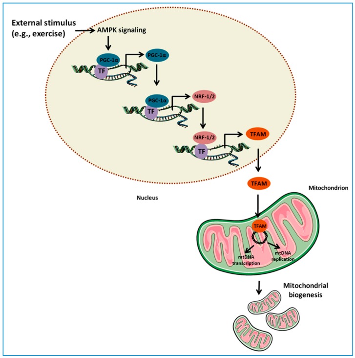
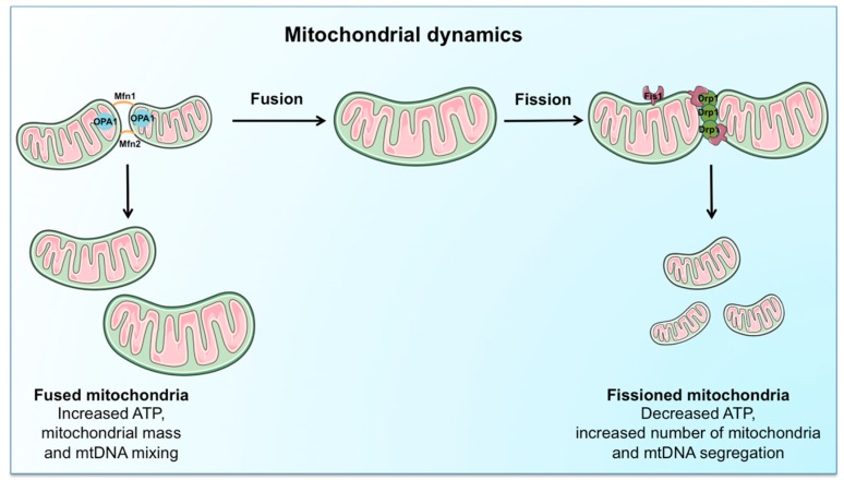
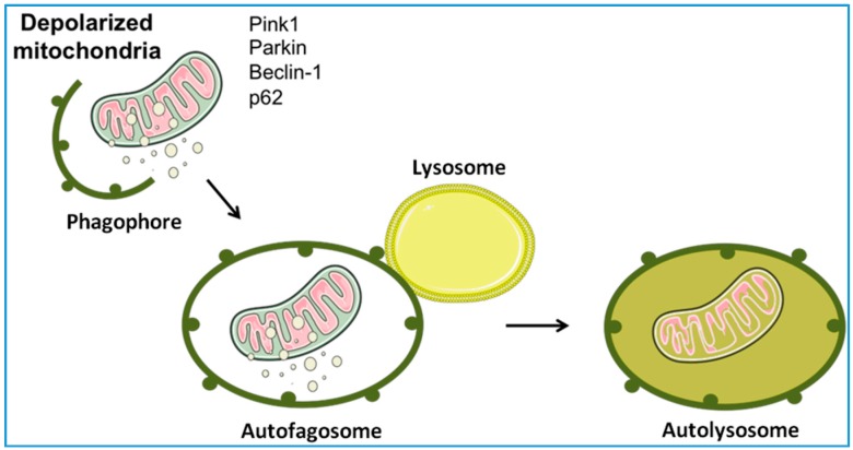
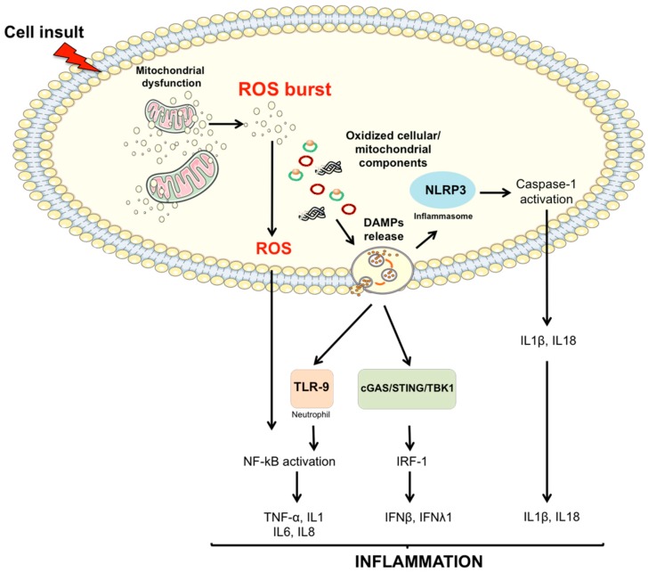
Among the complex determinants of aging, mitochondrial dysfunction has been in the spotlight for a long time. As the hub for many cellular functions, the maintenance of an adequate pool of functional mitochondria is crucial for tissue homeostasis. Their unique role in energy supply makes these organelles essential, especially in those tissues strictly dependent on oxidative metabolism. Mitochondrial quality control (MQC) is ensured by pathways related to protein folding and degradation as well as by processes involving the entire organelle, such as biogenesis, dynamics, and mitophagy. Dysfunctional MQC, oxidative stress and inflammation are hallmarks of senescence and chronic degenerative diseases. One of the consequences of age-related failing MQC and oxidative stress is the release of mitochondria-derived damage-associated molecular patterns (DAMPs). Through their bacterial ancestry, these molecules contribute to mounting an inflammatory response by interacting with receptors similar to those involved in pathogen-associated responses. Mitochondrial DAMPs, especially cell-free mitochondrial DNA, have recently become the subject of intensive research because of their possible involvement in conditions associated with inflammation, such as aging and degenerative diseases. Here, we review the contribution of mitochondrial DAMPs to inflammation and discuss some of the mechanisms at the basis of their generation.

Keywords: TFAM; damage-associated molecular patterns (DAMPs); inflammasome; mitochondrial biogenesis; mitochondrial dynamics; mitochondrial quality control (MQC); mitophagy; sterile inflammation.


Conflict of interest statement

Emanuele Marzetti, Francesco Landi, Roberto Bernabei, and Riccardo Calvani are partners of the SPRINTT consortium, which is partly funded by the European Federation of Pharmaceutical Industries and Associations (EFPIA). Emanuele Marzetti served as a consultant for Huron Consulting Group, Genactis, and Novartis; Riccardo Calvani served as a consultant from Novartis. The other authors declare no conflict of interest.

Figures

Figure 1
Figure 1
Schematic representation of the regulation of mitochondrial biogenesis. In response to external stimuli (e.g., skeletal muscle contraction or exercise), the nuclear genome coordinates the expression of nuclear and mitochondrial proteins. This pathway is triggered by the activation of signaling molecules, including AMP-activated protein kinase (AMPK), and converges on the expression of the transcriptional coactivator peroxisome proliferator-activated receptor gamma coactivator-1α (PGC-1α), the master regulator of mitochondrial biogenesis. PGC-1α promotes its own expression as well as that of the nuclear respiratory factor 1 and 2 (NRF-1/2). NRF-1 and 2 bind and up-regulate the expression of nuclear genes encoding mitochondrial proteins as well as the expression of mitochondrial transcription factor A (TFAM), which is subsequently transported into mitochondria. Here, TFAM binds to mitochondrial DNA (mtDNA) and activates the transcription and replication of the mitochondrial genome, a crucial step in the generation of new organelles. TF, transcription factor.
Figure 2
Figure 2
Schematic representation of the regulation of mitochondrial dynamics. The coordination of fusion and fission events is ensured by a complex machinery, involving mitofusin (Mfn) 1 and 2, optic atrophy protein 1 (OPA1), dynamin-related protein 1 (Drp1), and fission protein 1 (Fis1). Mitochondrial fusion, by interconnecting organelles, promotes mtDNA mixing and enhances bioenergetic efficiency. Mitochondrial fission, instead, ensures equal organelle segregation between daughter cells, reduces ATP generation, and targets defective mitochondria for their subsequent disposal via mitophagy.
Figure 3
Figure 3
Schematic representation of mitophagy. Mitophagy ensures the selective degradation of dysfunctional mitochondria through specialized autophagy. The process begins with the formation of a double-layered membrane (phagophore) around the organelles to be degraded. By growing in size, the phagophore progressively engulfs the cargo, forming an autophagosome. The subsequent autophagosome fusion with lysosomes generates an autolysosome wherein the cargo is digested. PINK1, PTEN-induced putative kinase 1.
Figure 4
Figure 4
Proposed signaling pathways through which damaged-associated molecular patterns (DAMPs) can trigger inflammation. The impairment of mitochondrial quality control processes may lead to the accumulation of intracellular oxidized components and their release as DAMPs. Damaged mtDNA molecules, either TFAM-bound (green circles) or unbound (red circles) may be released as DAMPs. These, in turn, can activate an inflammatory response via three distinct signaling pathways by interacting with (1) Toll-like receptors (TLRs), (2) nucleotide-binding oligomerization domain (NOD)-like receptor family pyrin domain containing 3 (NLRP3) inflammasome, and (3) cytosolic cyclic GMP-AMP synthase (cGAS)-stimulator of interferon genes (STING) DNA-sensing system. IFN, interferon; IL, interleukin; IRF-1, interferon regulatory factor 1; mtDNA, mitochondrial DNA; NF-κB, nuclear factor κB; ROS, reactive oxygen species; TBK1, TANK-binding kinase 1; TNF-α, tumor necrosis factor α.

References

    1. Seals D.R., Justice J.N., LaRocca T.J. Physiological geroscience: Targeting function to increase healthspan and achieve optimal longevity. J. Physiol. 2016;594:2001–2024. doi: 10.1113/jphysiol.2014.282665. - DOI - PMC - PubMed
    1. Harman D. Free radical theory of aging: Consequences of mitochondrial aging. Age. 1983;6:86–94. doi: 10.1007/BF02432509. - DOI
    1. Miquel J., Economos A.C., Fleming J., Johnson J.E., Jr. Mitochondrial role in cell aging. Exp. Gerontol. 1980;15:575–591. doi: 10.1016/0531-5565(80)90010-8. - DOI - PubMed
    1. Ajioka R.S., Phillips J.D., Kushner J.P. Biosynthesis of heme in mammals. Biochim. Biophys. Acta. 2006;1763:723–736. doi: 10.1016/j.bbamcr.2006.05.005. - DOI - PubMed
    1. De Stefani D., Rizzuto R., Pozzan T. Enjoy the trip: Calcium in mitochondria back and forth. Annu. Rev. Biochem. 2016;85:161–192. doi: 10.1146/annurev-biochem-060614-034216. - DOI - PubMed