. 2017 Jun 21;28(6):1767-1776.
doi: 10.1021/acs.bioconjchem.7b00237. Epub 2017 May 18.

Intraperitoneal Administration of Neural Stem Cell-Nanoparticle Conjugates Targets Chemotherapy to Ovarian Tumors

Affiliations

Intraperitoneal Administration of Neural Stem Cell-Nanoparticle Conjugates Targets Chemotherapy to Ovarian Tumors

Pengpeng Cao et al. Bioconjug Chem. .

Abstract

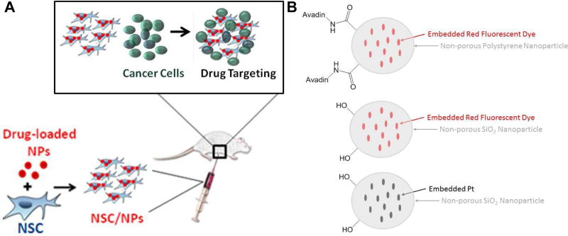
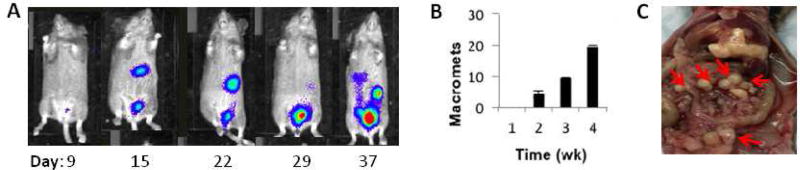
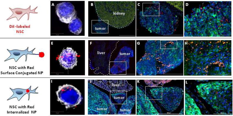
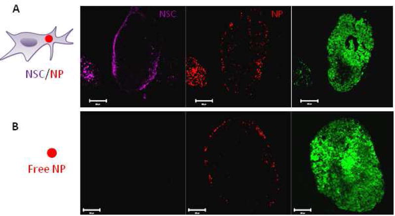
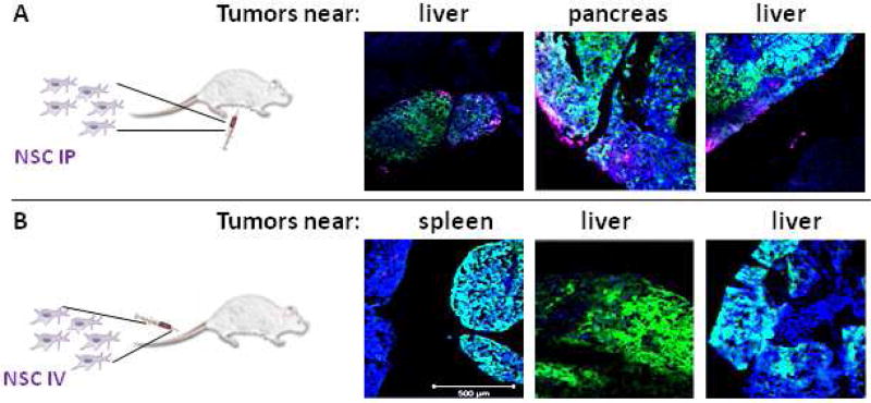
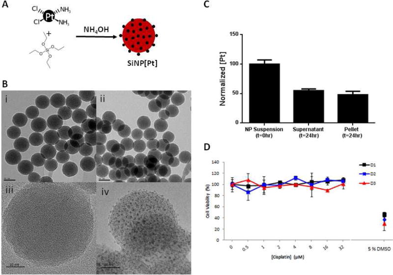
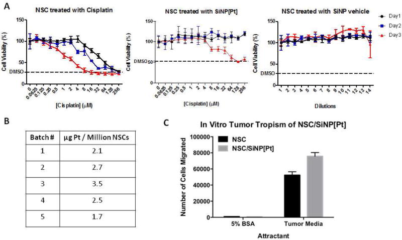
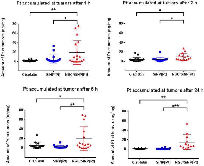
Ovarian cancer is particularly aggressive once it has metastasized to the abdominal cavity (stage III). Intraperitoneal (IP) as compared to intravenous (IV) administration of chemotherapy improves survival for stage III ovarian cancer, demonstrating that concentrating chemotherapy at tumor sites has therapeutic benefit; unfortunately, IP therapy also increases toxic side effects, thus preventing its completion in many patients. The ability to target chemotherapy selectively to ovarian tumors while sparing normal tissue would improve efficacy and decrease toxicities. We have previously shown that tumor-tropic neural stem cells (NSCs) dramatically improve the intratumoral distribution of nanoparticles (NPs) when given intracerebrally near an orthotopic brain tumor or into a flank xenograft tumor. Here, we show that NPs either conjugated to the surface of NSCs or loaded within the cells are selectively delivered to and distributed within ovarian tumors in the abdominal cavity following IP injection, with no evidence of localization to normal tissue. IP administration is significantly more effective than IV administration, and NPs carried by NSCs show substantially deeper penetration into tumors than free NPs. The NSCs and NPs target and localize to ovarian tumors within 1 h of administration. Pt-loaded silica NPs (SiNP[Pt]) were developed that can be transported in NSCs, and it was found that the NSC delivery of SiNP[Pt] (NSC-SiNP[Pt]) results in higher levels of Pt in tumors as compared to free drug or SiNP[Pt]. To the best of our knowledge, this work represents the first demonstration that cells given IP can target the delivery of drug-loaded NPs.


Conflict of interest statement

The remaining authors declare no conflict of interest.

Figures

Figure 1
Figure 1
(A) Schematic of desired targeting. (B) Particles used in this manuscript, note only two surface functional groups are shown for each particle type. In reality there are many surface functional groups.
Figure 2
Figure 2. OVCAR8 ovarian cancer model
(A) Mice injected IP with 2e6 OVCAR8.ffluc cells developed widespread metastatic disease in the abdomen. (B) Tumor progression over 4 weeks observed in n=3 mice/time point. Micrometastases are observed by week 1. Macrometastases are first seen at week 2, and increase in number, size and distribution over time. Data represents average number of macroscopic metastases +/− SEM. (C) Organ pluck at 3 weeks showing ovarian metastases (small white nodules; red arrows) on liver, pancreas and intestinal surfaces.
Figure 3
Figure 3. Neural stem cell tropism to peritoneal ovarian cancer metastasis
(A,E,I) Representative confocal z-stack images of native neural stem cells (A), and two different NSC/NP constructs in which the NPs are either localized to the cell surface (E) or internalized (I). Cell nuclei and cytoskeleton are respectively visualized using DAPI (blue) and phalloidin staining (pseudo-colored white). Tumor cells expressed GFP (green). Note: The kidney has a very strong background signal when visualizing GFP. (B–D) NSCs labeled with CellTracker CM-DiI demonstrate good distribution in tumor but not in adjacent normal kidney (2 million NSC.DiI in 200 µL PBS injected IP on Day 38; then harvested 4 days post-NSC injection). (F–H) NSCs with surface conjugated NPs demonstrate good distribution in tumor but not adjacent normal liver (4 million NSC.NP in 1 mL PBS injected IP on Day 13: harvested 4 days post-NSC injection). (J–I) NSCs with internalized NPs demonstrate good distribution in tumor but not adjacent normal liver (4 million NSC/NPs in 1 mL PBS injected IP on Day 25: harvested 4 days post-NSC injection). Scale bars: B,F,J: 1000 microns; C,G,K: 500 microns; D,H,L: 200 microns.
Figure 4
Figure 4. NSC-delivery improves silica nanoparticle penetration
(A–B) Representative fluorescent images of tumor sections collected 4 days after IP administration of 2 million NSC/SiNPs or free SiNPs on Day 21. (A) Isolated Far Red, and red channels demonstrate NSC/SiNPs exhibit excellent penetration of peritoneal tumors. Tumor signal (eGFP) is visible in the merged image. (B) Isolated Far Red, and red channels demonstrate SiNPs only surface-localized distribution at peritoneal tumors. Scale bars: 200 microns.
Figure 5
Figure 5. Neural stem cell administration route to peritoneal ovarian cancer metastasis
NSCs labeled with CellMask Deep Red plasma membrane stain (magenta) demonstrate good distribution when (A) administered IP but not when (B) administered IV in a mouse model of peritoneal ovarian metastasis established using OVCAR8.eGFP.ffluc cells. (2 million NSC.CellMask in 200 µL PBS injected IP on Day 21; then harvested 4 days post-NSC injection). Scale bars = 500 microns.
Figure 6
Figure 6
Preparation of Pt-loaded SiNPs. (A) Synthetic scheme (B) TEM and high resolution TEM imaging of control SiNPs (i, iii) and Pt-loaded SiNPs (ii, iv), (Scale bar = 50 nm in (i, ii), and 10 nm in (iii. iv). (C) After 24 hrs preincubation in PBS, 48 % of the Pt remains stably bound in the SiNPs (pellet) (D) Pt released from the SiNPs after 24 hrs (supernatant in C) showed no toxicity on the NSCs up to 72 hrs after treatment.
Figure 7
Figure 7
NSC can be loaded with NP[Pt] without compromising viability or migration A) SiNPs show no toxicity and SiNP[Pt] show no toxicity for 2 days B) NSCs can be reliably loaded with SiNP[Pt] C) loaded NSCs have unchanged migration in vitro.
Figure 8
Figure 8
NSC/SiNP[Pt] given IP enhance the retention of Pt at tumor sites. At 1, 2, 6 and 24 hours, tumors from NSC/SiNP[Pt]-treated animals had higher levels of Pt than those treated with free cisplatin or drug-loaded NPs. Statistical significance was determined using a two-tailed students t-test (* p <0.05, ** p<0.01, *** p<0.001).

References

    1. Lengyel E. Ovarian cancer development and metastasis. The American journal of pathology. 2010;177:1053–64. - PMC - PubMed
    1. Armstrong DK, Bundy B, Wenzel L, Huang HQ, Baergen R, Lele S, Copeland LJ, Walker JL, Burger RA, Gynecologic Oncology G. Intraperitoneal cisplatin and paclitaxel in ovarian cancer. N Engl J Med. 2006;354:34–43. - PubMed
    1. Aboody KS, Najbauer J, Metz MZ, D’Apuzzo M, Gutova M, Annala AJ, Synold TW, Couture LA, Blanchard S, Moats RA, et al. Neural Stem Cell–Mediated Enzyme/Prodrug Therapy for Glioma: Preclinical Studies. Science translational medicine. 2013;5:184ra59. - PMC - PubMed
    1. Aboody KS, Najbauer J, Danks MK. Stem and progenitor cell-mediated tumor selective gene therapy. Gene therapy. 2008;15:739–52. - PubMed
    1. Zhao D, Najbauer J, Annala AJ, Garcia E, Metz MZ, Gutova M, Polewski MD, Gilchrist M, Glackin CA, Kim SU, et al. Human neural stem cell tropism to metastatic breast cancer. Stem cells. 2012;30:314–25. - PubMed

Publication types

MeSH terms

Substances