. 2017 Apr 28;12(4):e0176693.
doi: 10.1371/journal.pone.0176693. eCollection 2017.

An efficient and robust MRI-guided radiotherapy planning approach for targeting abdominal organs and tumours in the mouse

Affiliations

An efficient and robust MRI-guided radiotherapy planning approach for targeting abdominal organs and tumours in the mouse

Veerle Kersemans et al. PLoS One. .

Abstract

Introduction: Preclinical CT-guided radiotherapy platforms are increasingly used but the CT images are characterized by poor soft tissue contrast. The aim of this study was to develop a robust and accurate method of MRI-guided radiotherapy (MR-IGRT) delivery to abdominal targets in the mouse.

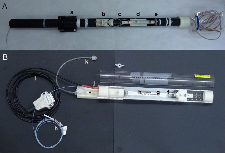
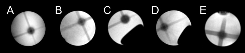
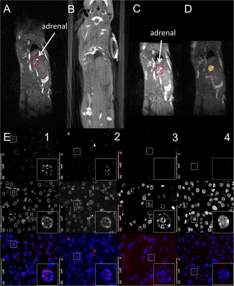
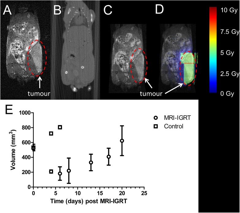
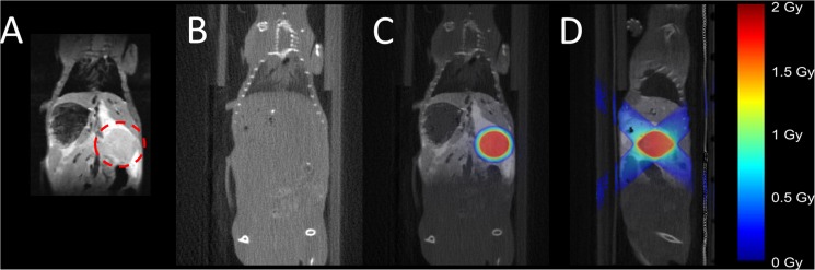
Methods: A multimodality cradle was developed for providing subject immobilisation and its performance was evaluated. Whilst CT was still used for dose calculations, target identification was based on MRI. Each step of the radiotherapy planning procedure was validated initially in vitro using BANG gel dosimeters. Subsequently, MR-IGRT of normal adrenal glands with a size-matched collimated beam was performed. Additionally, the SK-N-SH neuroblastoma xenograft model and the transgenic KPC model of pancreatic ductal adenocarcinoma were used to demonstrate the applicability of our methods for the accurate delivery of radiation to CT-invisible abdominal tumours.

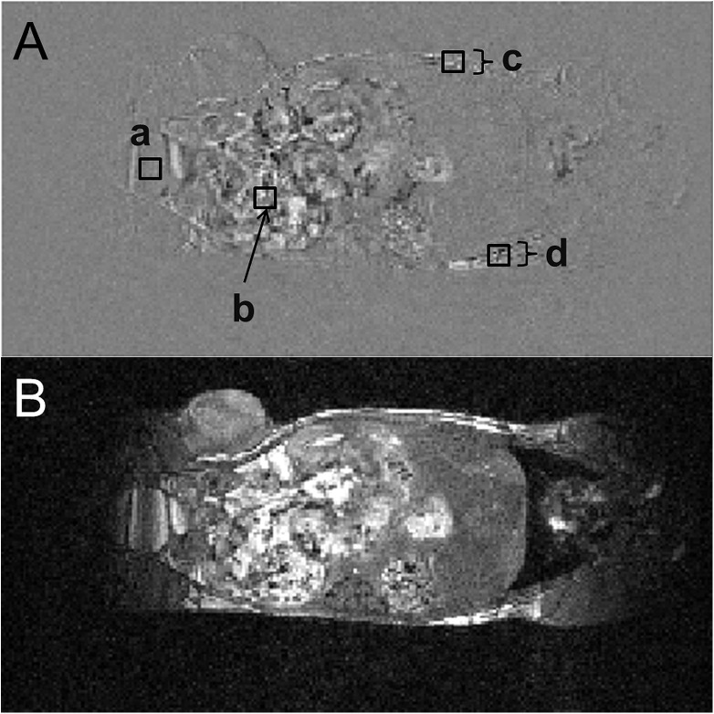
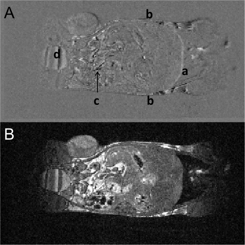
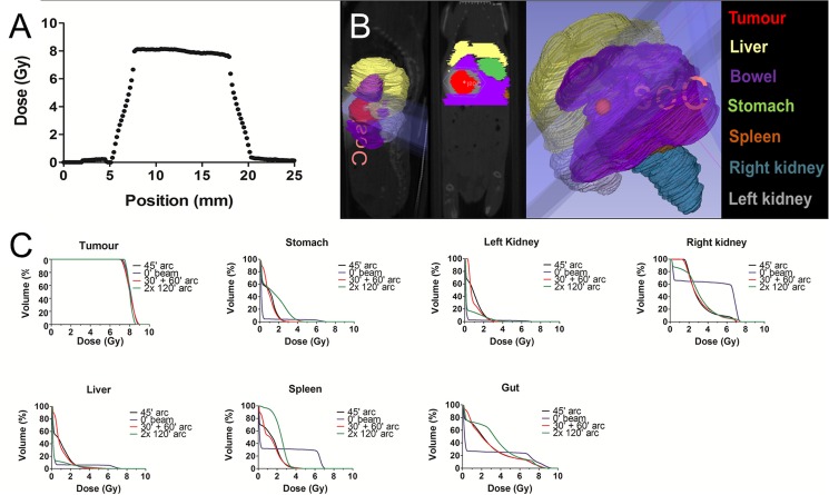
Results: The BANG gel phantoms demonstrated a targeting efficiency error of 0.56 ± 0.18 mm. The in vivo stability tests of body motion during MR-IGRT and the associated cradle transfer showed that the residual body movements are within this MR-IGRT targeting error. Accurate MR-IGRT of the normal adrenal glands with a size-matched collimated beam was confirmed by γH2AX staining. Regression in tumour volume was observed almost immediately post MR-IGRT in the neuroblastoma model, further demonstrating accuracy of x-ray delivery. Finally, MR-IGRT in the KPC model facilitated precise contouring and comparison of different treatment plans and radiotherapy dose distributions not only to the intra-abdominal tumour but also to the organs at risk.

Conclusion: This is, to our knowledge, the first study to demonstrate preclinical MR-IGRT in intra-abdominal organs. The proposed MR-IGRT method presents a state-of-the-art solution to enabling robust, accurate and efficient targeting of extracranial organs in the mouse and can operate with a sufficiently high throughput to allow fractionated treatments to be given.


Conflict of interest statement

Competing Interests: The authors have declared that no competing interests exist.

Figures

Fig 1
Fig 1. Photograph of the animal support cradle.
(A) Animal support cradle mounted in the MRI cradle assembly showing (a) MRI support cradle, (b) mounting block for the optical fibres for respiration monitoring, (c) optical fibres for respiration monitoring, (d) trapezoidal base of the animal support cradle which mated with a support block on the MRI support cradle, (e) RF coil. (B) Close-up of animal support cradle showing (f) mouthpiece assembly consisting of a base block, a vertical and horizontal adjustable mouth bar and an anaesthetic gas delivery tube, (g) electrical heating blanket, (h) optical fibre for temperature monitoring, (i) top of the animal support cradle with access points for the optical fibres for respiration monitoring, (j) quick-fit connector for electrical heating pad, (k) anaesthetic gas delivery tube with quick-fit connector and (l) power cable for the electrical heat delivery.
Fig 2
Fig 2. MR-IGRT of a painted feature in a BANG gel.
(A)—(E): Transversal view of MR-IGRT for the 5 tested BANG gels. A target volume was created by a pair of orthogonal 2 mm diameter (A, B, C, D) or 4 mm diameter (E) circular beams (volume 4.2 or 33.5 mm3) and targeted following MR-IGRT using a conical arc, delivered at a 45 degree oblique angle and using a 5 mm round collimator.
Fig 3
Fig 3. In vivo stability of body position: evaluation of intrinsic motions.
(A) Intrinsic motion as shown by taking the difference of the first and last image of a 30 minute acquisition. Bladder filling (a), gastrointestinal motility (b) and a general gravity induced body droop of up to 4 pixels (1 mm) (c, d) can be observed. The differences in signal intensity, expressed as the percentage of the first image, are 8%, 51%, 3% and 3% for regions a, b, c and d, respectively. (B) The corresponding anatomical image is shown.
Fig 4
Fig 4. In vivo stability of body position: evaluation of extrinsic motions.
(A) This represents the worst case image for extrinsic motion as shown by taking the difference image of the MR images produced before and after 4 cycles of transfer of the mouse in the cradle between the MR- and IGRT systems. Low level distortions can be observed at the diaphragm (a) and at the surface (b) of the mouse. Gastrointestinal motility (c) and bladder filling can also be observed (d). (B) The corresponding anatomical image is shown.
Fig 5
Fig 5. In vivo MR-IGRT of the adrenal glands and histological validation by p-γH2AXSer139 staining.
(A) respiratory-gated bSSFP MR image; (B): RT planning CBCT image; (C) CBCT-MR image to guide the RT treatment delivery; (D) treatment plan for the adrenal; (E) The top row shows fluorescent images of p-γH2AXSer139 that were acquired for 2 regions within the irradiated adrenal (column 1 and 2), the proximal kidney area to the irradiated adrenal (column 3) and the non-irradiated contralateral adrenal (column 4). The sections were counterstained with DAPI to visualize nucleus (middle row) and the merged image is shown in the bottom row. Representative images are shown.
Fig 6
Fig 6. In vivo MR-IGRT of neuroblastoma xenografts.
(A) respiratory-gated bSSFP MR image; (B): RT planning CBCT image; (C) CBCT-MR image to guide the RT treatment delivery; (D) dose distribution for the targeted treatment plan for a large tumour based on CBCT image to guide the RT treatment delivery: 2 radiotherapy fields with minimal overlap were used to cover the whole tumour whilst ensuring minimal exposure of surrounding normal tissues. The tumour is indicated within a dashed line; (E) Tumour volume of MRI-IGRT treated (5 Gy; n = 5) and non-irradiated control mice (n = 3). Values are expressed as mean ± standard deviation.
Fig 7
Fig 7. In vivo MR-IGRT of spontaneous pancreatic tumours in KPC mice.
(A) respiratory-gated bSSFP MR image; (B) RT planning CBCT image; (C) coronal and (D) sagittal view showing dose distribution (prescribed 2 Gy) based on CBCT-MR image to guide the RT treatment delivery. The colour heat map indicates the RT doses.
Fig 8
Fig 8. Determination of the optimal RT plan for intra-abdominal tumours in a mouse model of PDAC using a 10 mm diameter circular field.
(A) Planned dose profile for a pancreatic tumour determined at the isocentre, perpendicular to the angle of rotation (0°) and perpendicular to the sagittal axis of the mouse. (B) Illustration of MR image-based delineated tumour and organs at risk on the sagittal (left panel), coronal (middle panel) and 3-dimensionally, as indicated. (C) Mean dose-volume histograms for a prescribed single dose of 8 Gy showing the dosimetric distribution of four radiotherapy plans (45° Arc, 0° beam, 30°+60° arc and 2x 120° gantry arcs, respectively) to the intra-abdominal pancreatic tumour and the organs at risk in the KPC mouse model, as indicated.

References

    1. Tillner F, Thute P, Butof R, Krause M, Enghardt W. Pre-clinical research in small animals using radiotherapy technology—a bidirectional translational approach. Z Med Phys. 2014;24(4):335–51. doi: 10.1016/j.zemedi.2014.07.004 - DOI - PubMed
    1. Baumann BC, Benci JL, Santoiemma PP, Chandrasekaran S, Hollander AB, Kao GD, et al. An integrated method for reproducible and accurate image-guided stereotactic cranial irradiation of brain tumors using the small animal radiation research platform. Transl Oncol. 2012;5(4):230–7. PubMed Central PMCID: PMCPMC3431032. - PMC - PubMed
    1. Singh M, Murriel CL, Johnson L. Genetically engineered mouse models: closing the gap between preclinical data and trial outcomes. Cancer Res. 2012;72(11):2695–700. doi: 10.1158/0008-5472.CAN-11-2786 - DOI - PubMed
    1. Bibby MC. Orthotopic models of cancer for preclinical drug evaluation: advantages and disadvantages. Eur J Cancer. 2004;40(6):852–7. doi: 10.1016/j.ejca.2003.11.021 - DOI - PubMed
    1. Teicher BA. Tumor models for efficacy determination. Mol Cancer Ther. 2006;5(10):2435–43. doi: 10.1158/1535-7163.MCT-06-0391 - DOI - PubMed

Publication types

MeSH terms