. 2017 Apr 28;12(4):e0176795.
doi: 10.1371/journal.pone.0176795. eCollection 2017.

Selective mitochondrial DNA degradation following double-strand breaks

Affiliations

Selective mitochondrial DNA degradation following double-strand breaks

Amandine Moretton et al. PLoS One. .

Abstract

Mitochondrial DNA (mtDNA) can undergo double-strand breaks (DSBs), caused by defective replication, or by various endogenous or exogenous sources, such as reactive oxygen species, chemotherapeutic agents or ionizing radiations. MtDNA encodes for proteins involved in ATP production, and maintenance of genome integrity following DSBs is thus of crucial importance. However, the mechanisms involved in mtDNA maintenance after DSBs remain unknown. In this study, we investigated the consequences of the production of mtDNA DSBs using a human inducible cell system expressing the restriction enzyme PstI targeted to mitochondria. Using this system, we could not find any support for DSB repair of mtDNA. Instead we observed a loss of the damaged mtDNA molecules and a severe decrease in mtDNA content. We demonstrate that none of the known mitochondrial nucleases are involved in the mtDNA degradation and that the DNA loss is not due to autophagy, mitophagy or apoptosis. Our study suggests that a still uncharacterized pathway for the targeted degradation of damaged mtDNA in a mitophagy/autophagy-independent manner is present in mitochondria, and might provide the main mechanism used by the cells to deal with DSBs.


Conflict of interest statement

Competing Interests: The authors have declared that no competing interests exist.

Figures

Fig 1
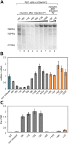
Fig 1. Loss of mtDNA after induction of the expression of a mitochondrial targeted PstI.
A. Map of the human mtDNA indicating the positions of the unique BamHI site and the two PstI sites, the distances between these sites and the positions of the probes used for the Southern blots (green asterisks). B. Southern blot analysis of the control HEK293 cells (WT) and stably transfected cells (PstI cells) after digestion by BamHI or BamHI + PstI for the control. PstI expression was induced for 2h with doxycycline and the cells were followed during the recovery period, from 0h to 2 days after induction. C. Quantification of the mtDNA/nDNA ratios in the PstI stably transfected cells, without induction or from 0h to 3 days after a 2h induction with doxycycline (mean ± s.e., N = 3). D. Changes in mitochondrial proteins levels during the recovery period after induction of PstI for 2h with doxycycline. Total cell lysates (20 μg) were analyzed by western blot with antibodies against mitochondrial (CO2: complex IV) or nuclear encoded subunits of the respiratory chain (ATP5a: complex V; UQCR2: complex III, SDHB: complex II; NDUFB8: complex I) and against DNA maintenance proteins, the helicase Twinkle, the DNA polymerase POLGA and TFAM. H3: Histone H3B (loading control).
Fig 2
Fig 2. Effect of silencing different mitochondrial DNA nucleases on the rapid mtDNA depletion.
A. Levels of different mitochondrial DNA nucleases before induction and during the recovery period after induction of PstI for 2h with doxycycline. Total cell lysates (20 μg) were analyzed by western blot with antibodies against ExoG, EndoG, MGME1, and FEN1. H3: Histone H3B (loading control). B. Southern blot analysis of the control HEK293 cells (WT) and stably transfected cells (PstI cells) after digestion by BamHI or BamHI + PstI for the control. The cells were grown in presence or absence of a mix of siRNAs targeting ExoG and/or EndoG. Three days after siRNA transfection, doxycycline was added to the cells for 2h to induce PstI expression, and the cell’s DNA was examined before induction and during the recovery period, at 0h, 5h and 1 day after induction. C, D, E. Similar experiments as in 2B, except that the siRNAs were targeting MGME1, DNA2 or FEN1, respectively.
Fig 3
Fig 3. The rapid loss of mtDNA is not correlated to a loss of mitochondria, autophagy or apoptosis.
Levels of A. mitochondrial proteins and C. an autophagy marker before induction and during the recovery period after induction of PstI for 2h with doxycycline. Total cell lysates (20 μg) were analyzed by western blot with antibodies against markers of the mitochondrial matrix (PDH), mitochondrial outer membrane (Tomm20), autophagy (LC3) and mitophagy (PINK1). CCCP is used as a positive control and Tubulin is used as loading control. Analysis of B. cell mitochondrial content and D. apoptosis by flow cytometry before induction and during the recovery period. * P ≤ 0.05 versus non-induced PstI cells for each cell population (Student’s t-test).
Fig 4
Fig 4. The loss of mtDNA is followed by a slow repopulation with intact mtDNA molecules.
A. Southern blot analysis of the control HEK293 cells (WT) and stably transfected cells (PstI cells) after digestion by BamHI or BamHI + PstI for the control. PstI expression was induced for 2h with doxycycline and cells were followed for 20 days. At day 20, a second induction of 2h with doxycycline was performed and the cells were followed during the second recovery period (marked with an apostrophe). B. Quantification of the mtDNA/nDNA ratios in the PstI stably transfected cells, from 0h to 41 days after a 2h induction with doxycycline and from 0h to 20days after a second 2h doxycycline induction. (mean ± s.e., N = 3). C. Quantification of the level of PstI transcripts in the control HEK293 cells (WT) and stably transfected cells (PstI cells), during 2 sequential inductions of PstI expression with doxycycline (mean ± s.e., N = 3). TBP is used as a reference RNA.

References

    1. Liang C, Ahmad K, Sue CM. The broadening spectrum of mitochondrial disease: Shifts in the diagnostic paradigm. Biochimica et Biophysica Acta (BBA)—General Subjects. 2014;1840(4):1360–1367. 10.1016/j.bbagen.2013.10.040 - DOI - PubMed
    1. McCrow JP, Petersen DC, Louw M, Chan EKF, Harmeyer K, Vecchiarelli S, et al. Spectrum of mitochondrial genomic variation and associated clinical presentation of prostate cancer in South African men. The Prostate. 2015;76(4):349–358. 10.1002/pros.23126 - DOI - PMC - PubMed
    1. Cha MY, Kim DK, Mook-Jung I. The role of mitochondrial DNA mutation on neurodegenerative diseases. Experimental & molecular medicine. 2015;47(3):e150 10.1038/emm.2014.122 - DOI - PMC - PubMed
    1. Herbst A, Wanagat J, Cheema N, Widjaja K, McKenzie D, Aiken JM. Latent mitochondrial DNA deletion mutations drive muscle fiber loss at old age. Aging Cell. 2016; 10.1111/acel.12520 - DOI - PMC - PubMed
    1. Milani L, Ghiselli F. Mitochondrial activity in gametes and transmission of viable mtDNA. Biology Direct. 2015;10(1). 10.1186/s13062-015-0057-6 - DOI - PMC - PubMed

LinkOut - more resources