. 2017 May 1;28(5):1005-1014.
doi: 10.1093/annonc/mdx047.

Epstein-Barr virus-associated gastric cancer reveals intratumoral heterogeneity of PIK3CA mutations

Affiliations

Epstein-Barr virus-associated gastric cancer reveals intratumoral heterogeneity of PIK3CA mutations

C Böger et al. Ann Oncol. .

Abstract

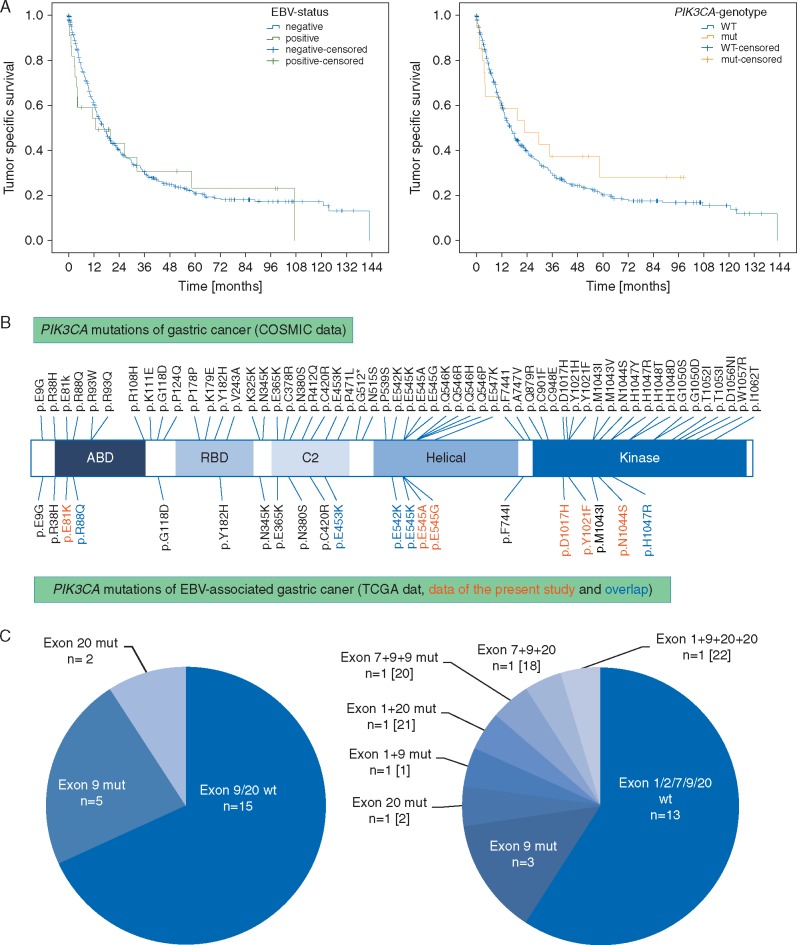
Background: Recent whole-genome sequencing identified four molecular subtypes of gastric cancer (GC), of which the subgroup of Epstein-Barr virus-associated GC (EBVaGC) showed a significant enrichment of PIK3CA mutations. We here aimed to validate independently the enrichment of PIK3CA mutations in EBVaGC of a Central European GC cohort, to correlate EBV status with clinico-pathological patient characteristics and to test for a major issue of GC, intratumoral heterogeneity.

Patients and methods: In a first step, 484 GCs were screened for EBV and PIK3CA hot spot mutations of exon 9/20 using EBER in situ hybridization and pyrosequencing, respectively. Secondly, an extended sequencing of PIK3CA also utilizing next generation sequencing was carried out in all EBVaGCs and 96 corresponding lymph node metastases.

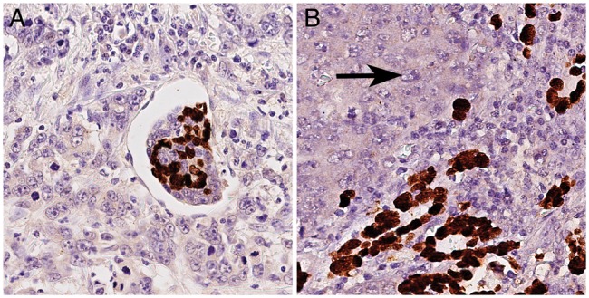
Results: Twenty-two GCs were EBER-positive, all being of latency type I. Intratumoral heterogeneity of EBER-positivity was found in 18% of EBVaGCs. Twenty-three GCs held PIK3CA mutations in hot spot regions of exon 9 or 20, being significantly more common in EBVaGCs (P < 0.001). Subsequent extended sequencing of PIK3CA of EBVaGCs showed that 14% harvested three to five different PIK3CA genotypes (including wildtype) in the same primary tumor, albeit in histologically and spatially distinct tumor areas, and that intratumoral heterogeneity of PIK3CA was also present in the corresponding lymph node metastases.

Conclusions: Our findings unravel issues of tumor heterogeneity and illustrate that the assessment of the EBV status in tissue biopsies might carry the risk of sampling errors, which may significantly hamper adequate molecular tumor classification in a more clinical setting. Moreover, this is the first report of intratumoral heterogeneity of PIK3CA mutations in GC, and our findings lead to the conclusion that PIK3CA mutant and -wildtype tumor subclones are skilled to metastasize independently to different regional lymph nodes.

Keywords: EBV; PI3K pathway; biomarker; intratumoral heterogeneity; lymph node metastases; next generation sequencing.


Figures

Figure 1.
Figure 1.
Study design. In a first step, the entire GC cohort (n = 484) was screened for EBV association and the presence of PIK3CA hot spot mutations of exon 9 and exon 20 using pyrosequencing. After validation of the significant enrichment of PIK3CA mutations in EBVaGCs, an extended PIK3CA mutational analysis was carried out for these cases in order to detect additional non-hot spot mutations. For 8 cases, only formalin fixed and paraffin embedded specimens were available, and Sanger sequencing of exon 1 and 9 and extended pyrosequencing of exon 20 was carried out. For 14 cases with available frozen, unfixed specimens (‘frozen’), an additional mutational analysis was done using the Illumina TruSight Tumor 26 panel on the Illumina MiSeq System.
Figure 2.
Figure 2.
Intratumoral heterogeneity of EBER-positivity. 4 of 22 EBVaGCs (18%; Table 2) showed intratumoral heterogeneity of EBER-positivity with a juxtaposition of EBER-negative and EBER-positive tumor areas. (A) illustrates the intermixture of EBER-positive and EBER-negative tumor cells within angioinvasion of a EBVaGC. EBER-heterogeneity was also observed within corresponding lymph node metastases (B; left: EBER-negative tumor cells (arrow); right: EBER-positive tumor cells). EBER-ISH, magnifications ×400.
Figure 3.
Figure 3.
Kaplan–Meier curves depicting patients’ survival according to EBV-infection status and PIK3CA mutational status (A). EBV- and PIK3CA-status had no significant effect on tumor-specific survival (P =0.712 for EBV; P =0.519 for PIK3CA). Comparison of COSMIC data with PIK3CA mutations of EBVaGC (B): This figure illustrates that PIK3CA mutations of EBVaGCs are distributed within all PIK3CA domains. Comparison of hot spot pyrosequencing versus NGS for the detection of PIK3CA mutations in EBVaGC (C): EBER-ISH detected 22 EBVaGCs, of which 7 cases held PIK3CA mutations in exon 9 or 20 after pyrosequencing of hot spot regions (left). The subsequent extended PIK3CA mutational analysis revealed that 5 EBVaGCs held three to five PIK3CA genotypes (including wildtype) and detected two additional PIK3CA mutated EBVaGCs (right). Both cases held PIK3CA mutations within the hot spot region of exon 9, which could be detected by pyrosequencing after minor modulation of the primer design. The numbers in brackets refer to the case number in Table 2.
Figure 4.
Figure 4.
Intratumoral heterogeneity of PIK3CA mutations in EBVaGC. Three cases held divergent mutations between corresponding FFPE and frozen specimens and two to four morphologically divergent tumor areas, which were subsequently sequenced anew separately. Case no. 22 had four morphologically distinct tumor areas on two different tumor slides, which were all EBV positive. On the first slide, area ‘1A’ accounted for 14% and was PIK3CA wildtype (blue color) and Her2/neu positive; area ‘1B’ accounted for 86% of the tumor section and was PIK3CA mutated in exon 9 (p.E545K; red color) and Her2/neu negative. On the second slide of the same case, area ‘2A’ accounted for 22% of the tumor section and was PIK3CA wildtype (blue color) and Her2/neu negative; area ‘2B’ accounted for 78%, had a mutation in exon 20 (p.N1044S; red color) and was Her2/neu negative. Specimen 1A and 1B showed divergent tumor morphology (1A: trabecular to solid growth pattern; 1B: glandular growth pattern) and a divergent PIK3CA mutational status. Specimen 1B and 2A showed concordant tumor morphology with a glandular growth pattern but divergent PIK3CA mutations. Magnifications: first line overviews; all other lines ×400.

Comment in

References

    1. Yuan DD, Zhu ZX, Zhang X, Liu J.. Targeted therapy for gastric cancer: current status and future directions. Oncol Rep 2016; 35: 1245–1254. - PubMed
    1. The Cancer Genome Atlas Research Network. Comprehensive molecular characterization of gastric adenocarcinoma. Nature 2014; 513: 202–209. - PMC - PubMed
    1. Böger C, Behrens HM, Mathiak M. et al. PD-L1 is an independent prognostic predictor in gastric cancer of Western patients. Oncotarget 2016; 7: 24269–24283. - PMC - PubMed
    1. Lin SJ, Gagnon-Bartsch JA, Tan IB. et al. Signatures of tumour immunity distinguish Asian and non-Asian gastric adenocarcinomas. Gut 2015; 64: 1721–1731. - PMC - PubMed
    1. Lauren T. The two histologic main types of gastric carcinoma: diffuse and so-called intestinal-type carcinoma. Acta Pathol Microbiol Scand 1965; 64: 31–49. - PubMed

MeSH terms

Substances