. 2017 May 1;19(5):699-709.
doi: 10.1093/neuonc/now254.

Targeted next-generation sequencing of pediatric neuro-oncology patients improves diagnosis, identifies pathogenic germline mutations, and directs targeted therapy

Affiliations

Targeted next-generation sequencing of pediatric neuro-oncology patients improves diagnosis, identifies pathogenic germline mutations, and directs targeted therapy

Cassie N Kline et al. Neuro Oncol. .

Erratum in

  • Corrigenda.
    [No authors listed] [No authors listed] Neuro Oncol. 2017 Apr 1;19(4):601. doi: 10.1093/neuonc/nox024. Neuro Oncol. 2017. PMID: 28388712 Free PMC article. No abstract available.

Abstract

Background: Molecular profiling is revolutionizing cancer diagnostics and leading to personalized therapeutic approaches. Herein we describe our clinical experience performing targeted sequencing for 31 pediatric neuro-oncology patients.

Methods: We sequenced 510 cancer-associated genes from tumor and peripheral blood to identify germline and somatic mutations, structural variants, and copy number changes.

Results: Genomic profiling was performed on 31 patients with tumors including 11 high-grade gliomas, 8 medulloblastomas, 6 low-grade gliomas, 1 embryonal tumor with multilayered rosettes, 1 pineoblastoma, 1 uveal ganglioneuroma, 1 choroid plexus carcinoma, 1 chordoma, and 1 high-grade neuroepithelial tumor. In 25 cases (81%), results impacted patient management by: (i) clarifying diagnosis, (ii) identifying pathogenic germline mutations, or (iii) detecting potentially targetable alterations. The pathologic diagnosis was amended after genomic profiling for 6 patients (19%), including a high-grade glioma to pilocytic astrocytoma, medulloblastoma to pineoblastoma, ependymoma to high-grade glioma, and medulloblastoma to CNS high-grade neuroepithelial tumor with BCOR alteration. Multiple patients had pathogenic germline mutations, many of which were previously unsuspected. Potentially targetable alterations were identified in 19 patients (61%). Additionally, novel likely pathogenic alterations were identified in 3 cases: an in-frame RAF1 fusion in a BRAF wild-type pleomorphic xanthoastrocytoma, an inactivating ASXL1 mutation in a histone H3 wild-type diffuse pontine glioma, and an in-frame deletion within exon 2 of MAP2K1 in a low-grade astrocytic neoplasm.

Conclusions: Our experience demonstrates the significant impact of molecular profiling on diagnosis and treatment of pediatric brain tumors and confirms its feasibility for use at the time of diagnosis or recurrence.

Keywords: molecular profiling; next-generation sequencing; pediatric brain tumors; personalized therapy; targeted therapeutics.


Figures

Fig. 1
Fig. 1
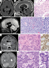
Genomic profiling improves diagnostic accuracy. (A–C) Tectal glioma in a 9-year-old boy initially diagnosed as astrocytoma of uncertain grade found to have KIAA1549-BRAF fusion leading to amended diagnosis of pilocytic astrocytoma, WHO grade I. (A) Axial T2-weighted FLAIR MR image revealing a tectal mass with extension into bilateral thalami. (B) Sagittal T1-weighted post-gadolinium MR image revealing peripheral enhancement and exophytic growth into the third ventricle with obstructive hydrocephalus. (C) Hematoxylin and eosin (H&E) stained section of the tumor. (D–F) Primitive neuroectodermal tumor in the midline of the posterior fossa of an 18-month-old boy initially diagnosed as medulloblastoma found to have somatic DICER1 mutation leading to amended diagnosis of pineoblastoma. (D) Sagittal T2-weighted FLAIR MR image revealing a circumscribed solid mass centered in the midline within the region of the pineal gland causing compression of the subjacent cerebellar vermis. (E–F) H&E stained sections of the tumor. (G–I) Suprasellar mass with cerebrospinal dissemination in an 11-year-old boy initially diagnosed as ependymoma found to have FGFR1 mutation leading to amended diagnosis of high-grade glioma. (G) Coronal T1-weighted post-gadolinium MR image revealing a complex, solid, and cystic mass in the suprasellar space. (H) H&E stained section of the tumor. (I) Immunohistochemistry showing diffuse strong staining for OLIG2 in tumor cells. (J–L) High-grade neoplasm in the cerebellum of a 4-year-old girl initially diagnosed as medulloblastoma found to have internal tandem duplication within exon 15 of BCOR leading to amended diagnosis of CNS high-grade neuroepithelial tumor with BCOR alteration. (J) Coronal T2-weighted MR image. (K–L) H&E stained sections of the tumor. Sequencing reads containing the BCOR internal tandem duplication are shown in Supplementary Fig. 3. Scale bar, 20 µm.
Fig. 2
Fig. 2
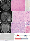
Genomic profiling identifies novel pathogenic germline mutations. (A–D) Medulloblastoma with anaplastic/large cell histologic features in a 5-year-old boy found to have monoallelic germline PALB2 nonsense mutation with loss of heterozygosity in the tumor. (A) Coronal T1-weighted post-gadolinium MR image revealing an enhancing multinodular mass centered in the fourth ventricle and left cerebellar hemisphere. (B–C) Hematoxylin and eosin (H&E) stained sections of the tumor. (D) Genome-wide copy number plot of the tumor showing numerous chromosomal gains and losses, typical of tumors with defects in homologous recombination. (E–I) Uveal ganglioneuroma and cerebral cavernous malformation in a 5-year-old girl found to have germline PTEN mutation with loss of heterozygosity in the tumor. (E) Axial T1-weighted MR image of the orbits revealing diffuse thickening of the uveal tract in the right globe. (F–H) H&E stained sections of the tumor showing a spindle cell neoplasm diffusely expanding the uveal tract including the ciliary body (F) and causing retinal atrophy and detachment (G). Large dysmorphic ganglion cells were admixed among the neoplastic spindle cells, diagnostic of ganglioneuroma (H). (I) Coronal T2-weighted FLAIR MR image revealing a cavernous malformation in the left occipital lobe. Scale bar, 20 µm.
Fig. 3
Fig. 3
Genomic profiling identifies novel likely pathogenic alterations. (A–C) Diffuse intrinsic pontine glioma in a 12-year-old boy lacking histone H3 mutation found to have a somatic inactivating mutation in ASXL1. (A) Axial T2-weighted FLAIR MR image. (B) Hematoxylin and eosin (H&E) stained section of the tumor showing an infiltrating astrocytoma without high-grade features. (C) Immunohistochemistry showing absence of staining for histone H3-K27M mutant protein. (D–G, K) Low-grade astrocytic neoplasm in a 12-year-old boy found to have small in-frame deletion within exon 2 of MAP2K1. (D) Coronal T2-weighted FLAIR MR image showing an ill-defined, expansile mass in the left medial temporal lobe. (E–F) H&E stained sections showing a low-grade astrocytic neoplasm with densely fibrillary background. (K) Sequencing reads for the tumor mapping to exon 2 of MAP2K1 with many containing an in-frame 15 bp deletion. (G–J, L) Recurrent pleomorphic xanthoastrocytoma with anaplastic features in a 19-year-old man lacking BRAF mutation found to have ATG7-RAF1 fusion. (G) Coronal T2-weighted FLAIR MR image showing a circumscribed mass in the superficial cortex of the left parietal lobe. (H) H&E stained section showing a solid neoplasm of pleomorphic astrocytes. (I) Periodic acid–Schiff stain showing one of the many eosinophilic granular bodies in the tumor. (J) Laidlaw reticulin stain demonstrating intercellular basement membrane deposition by the neoplastic astrocytes. (L) Genetic diagram of the ATG7 and RAF1 loci on chromosome 3, along with the inv(3)(p25.3;p25.2) identified in the tumor resulting in production of an in-frame fusion between exons 1–18 of ATG7 and exons 8–17 of RAF1 encoding the serine/threonine kinase domain. Scale bar, 20 µm.

References

    1. Broniscer A, Gajjar A. Supratentorial high-grade astrocytoma and diffuse brainstem glioma: two challenges for the pediatric oncologist. Oncologist. 2004;9(2):197–206. - PubMed
    1. Northcott PA, Pfister SM, Jones DT. Next-generation (epi)genetic drivers of childhood brain tumours and the outlook for targeted therapies. Lancet Oncol. 2015;16(6):e293–e302. - PubMed
    1. Nikiforova MN, Wald AI, Melan MA, et al. Targeted next-generation sequencing panel (GlioSeq) provides comprehensive genetic profiling of central nervous system tumors. Neuro Oncol. 2016;18(3):379–387. - PMC - PubMed
    1. Sahm F, Schrimpf D, Jones DT, et al. Next-generation sequencing in routine brain tumor diagnostics enables an integrated diagnosis and identifies actionable targets. Acta Neuropathol. 2016;131(6):903–910. - PubMed
    1. Hoffman LM, Salloum R, Fouladi M. Molecular biology of pediatric brain tumors and impact on novel therapies. Curr Neurol Neurosci Rep. 2015;15(4):10. - PubMed