. 2017 May 1;19(5):719-725.
doi: 10.1093/neuonc/now194.

Incidence of pseudoprogression in low-grade gliomas treated with radiotherapy

Affiliations

Incidence of pseudoprogression in low-grade gliomas treated with radiotherapy

Sophie E van West et al. Neuro Oncol. .

Abstract

Background: As the incidence of pseudo-progressive disease (psPD), or pseudoprogression, in low-grade glioma (LGG) is unknown, we retrospectively investigated this phenomenon in a cohort of LGG patients given radiotherapy (RT).

Methods: All MRI scans and clinical data from patients with histologically proven LGG treated with radiation between 2000 and 2011 were reviewed. PsPD was scored when a new enhancing lesion occurred after RT and subsequently disappeared or remained stable for at least a year without therapy, including dexamethasone.

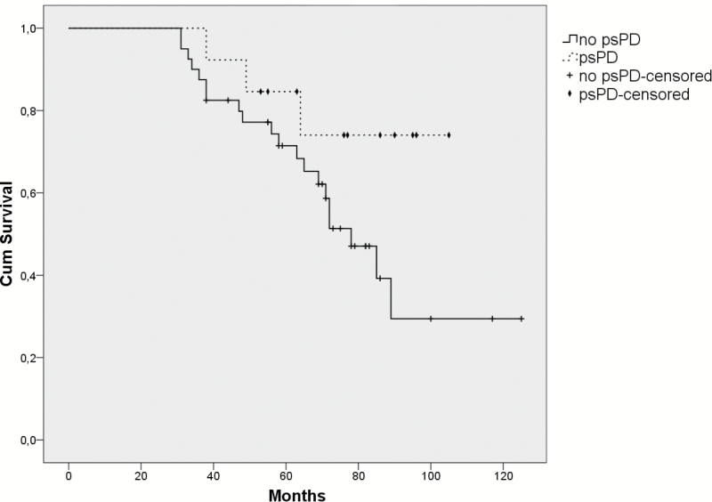
Results: Sixty-three out of 71 patients who received RT for LGG were deemed eligible for evaluation of psPD. The median follow-up was 5 years (range 1‒10 y). PsPD was seen in 13 patients (20.6%). PsPD occurred after a median of 12 months with a range of 3-78 months. The median duration of psPD was 6 months, with a range of 2-26 months and always occurred within the RT high dose fields of at least 45 Gy. The area of the enhancement at the time of psPD was significantly smaller compared with the area of enhancement during "true" progression (median size 54mm2 [range 12-340mm2] vs 270mm2 [range 30-3420mm2], respectively; P = .009).

Conclusions: PsPD occurs frequently in LGG patients receiving RT. This supports the policy to postpone a new line of treatment until progression is evident, especially when patients have small contrast enhancing lesions within the RT field.

Keywords: LGG; glioma; low-grade glioma; pseudoprogression; radiotherapy.


Figures

Fig. 1.
Fig. 1.
MRI scans from 32-year-old male with an astrocytoma in the right frontal-temporal lobe, treated with postoperative radiation (total dose 50.4 Gy). (A) MRI scan 36 months after the RT with no enhancement. (B) MRI scan 42 months after RT, with an enhancing lesion in the ventricle wall of the opposite left hemisphere, but still within the RT field. (C) MRI scan 45 months after RT and 3 months after the appearance of the enhancing lesion shows no enhancement. He received no dexamethasone or other treatments during this period and remained clinically stable. He radiologically progressed 53 months after the RT.
Fig. 2.
Fig. 2.
MRI scans from 53-year-old female with an oligoastrocytoma treated with postoperative radiation (total dose 60 Gy). (A) MRI scan 7 months after the RT with no enhancement. (B) MRI scan 14 months after RT with an enhancing lesion in the left ventricle wall (arrow), within the RT field. (C) MRI scan 21 months after RT and 14 months after the appearance of the enhancing lesion shows no enhancement. The patient remained clinically stable in this period without need for dexamethasone and without further treatments. At the time of analysis for this study, 84 months after the RT, she had no progressive disease.
Fig. 3.
Fig. 3.
MRI scans from 46-year-old male with an oligodendroglioma in the right frontal-temporal lobe, treated with postoperative radiation (total dose 50.4 Gy). (A) MRI scan 22 months after the RT with no enhancement. (B) MRI scan 25 months after RT shows an enhancing lesion in the right temporal lobe, within the RT field. (C) MRI scan 41 months after RT, and 26 months after the appearance of the enhancing lesion it has disappeared. No dexamethasone or further treatments were given and he remained clinically stable. At the time of analysis for this study, 43 months after the RT, he still had no progressive disease.
Fig. 4.
Fig. 4.
The Kaplan–Meier survival curves of the patients with psPD versus the patients without psPD. Patients without psPD were excluded from this analysis if they died before the 95th percentile of the time to development of psPD (30.8 mo), to avoid a possible time bias, as patients with psPD have to live long enough to meet the definition of psPD. After this correction, the OS between the patients with and without psPD was not significantly different (log-rank, P = .090).

References

    1. van den Bent MJ, Wefel JS, Schiff D, et al. Response assessment in neuro-oncology (a report of the RANO group): assessment of outcome in trials of diffuse low-grade gliomas. Lancet Oncol. 2011;12(6):583–593. - PubMed
    1. Kumthekar P, Raizer J, Singh S. Low-grade glioma. Cancer Treat Res. 2015;163:75–87. - PubMed
    1. Claus EB, Walsh KM, Wiencke JK, et al. Survival and low-grade glioma: the emergence of genetic information. Neurosurg Focus. 2015;38(1):E6. - PMC - PubMed
    1. Buckner JC, Shaw EG, Pugh SL, et al. Radiation plus procarbazine, CCNU, and vincristine in low-grade glioma. N Engl J Med. 2016;374(14):1344–1355. - PMC - PubMed
    1. Rath JJ, Smits M, Ducray F, et al. Increased rCBV in status epilepticus. J Neurol. 2012;259(8):1746–1748. - PMC - PubMed

MeSH terms