. 2017 Oct 19;19(11):1494-1502.
doi: 10.1093/neuonc/nox085.

Detection of wild-type EGFR amplification and EGFRvIII mutation in CSF-derived extracellular vesicles of glioblastoma patients

Affiliations

Detection of wild-type EGFR amplification and EGFRvIII mutation in CSF-derived extracellular vesicles of glioblastoma patients

Javier M Figueroa et al. Neuro Oncol. .

Abstract

Background: RNAs within extracellular vesicles (EVs) have potential as diagnostic biomarkers for patients with cancer and are identified in a variety of biofluids. Glioblastomas (GBMs) release EVs containing RNA into cerebrospinal fluid (CSF). Here we describe a multi-institutional study of RNA extracted from CSF-derived EVs of GBM patients to detect the presence of tumor-associated amplifications and mutations in epidermal growth factor receptor (EGFR).

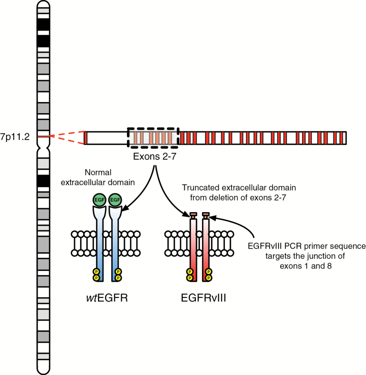
Methods: CSF and matching tumor tissue were obtained from patients undergoing resection of GBMs. We determined wild-type (wt)EGFR DNA copy number amplification, as well as wtEGFR and EGFR variant (v)III RNA expression in tumor samples. We also characterized wtEGFR and EGFRvIII RNA expression in CSF-derived EVs.

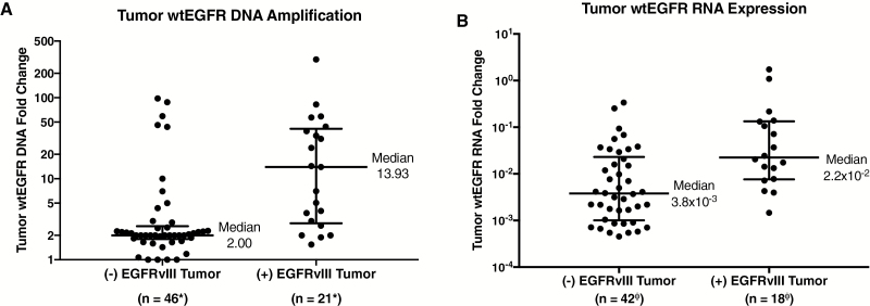
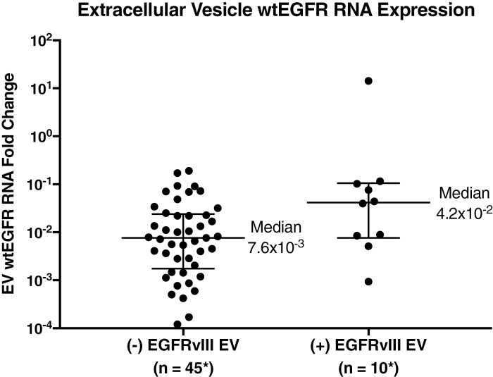
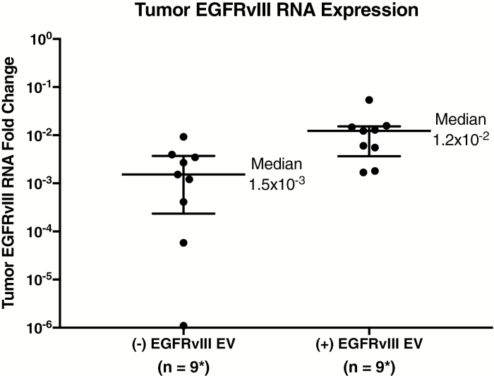
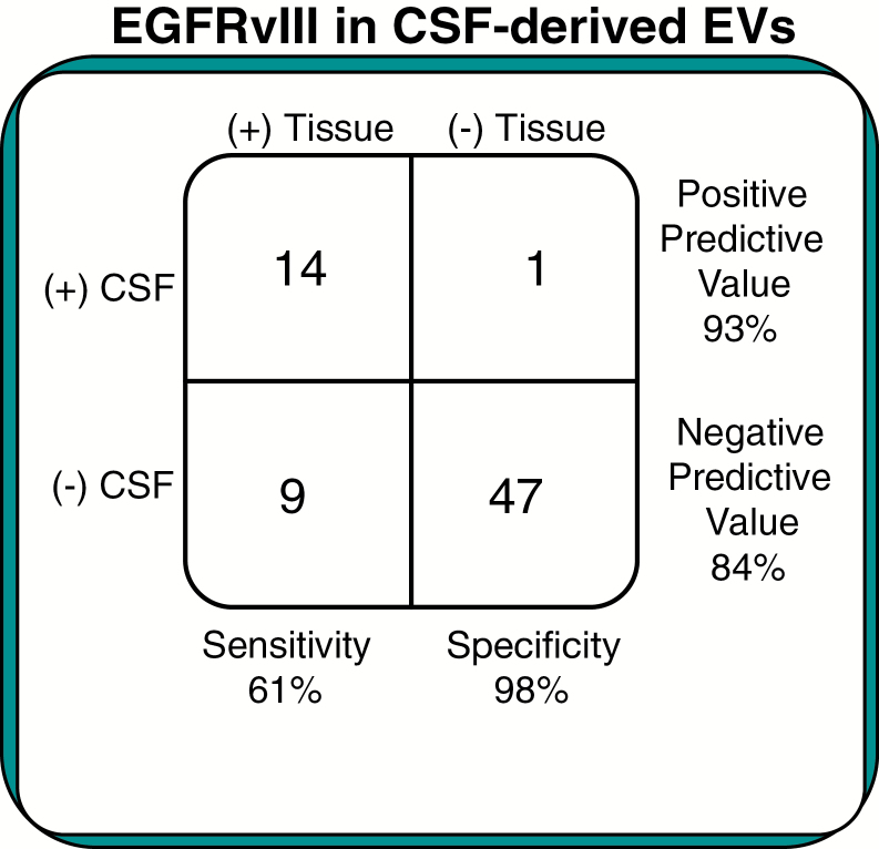
Results: EGFRvIII-positive tumors had significantly greater wtEGFR DNA amplification (P = 0.02) and RNA expression (P = 0.03), and EGFRvIII-positive CSF-derived EVs had significantly more wtEGFR RNA expression (P = 0.004). EGFRvIII was detected in CSF-derived EVs for 14 of the 23 EGFRvIII tissue-positive GBM patients. Conversely, only one of the 48 EGFRvIII tissue-negative patients had the EGFRvIII mutation detected in their CSF-derived EVs. These results yield a sensitivity of 61% and a specificity of 98% for the utility of CSF-derived EVs to detect an EGFRvIII-positive GBM.

Conclusion: Our results demonstrate CSF-derived EVs contain RNA signatures reflective of the underlying molecular genetic status of GBMs in terms of wtEGFR expression and EGFRvIII status. The high specificity of the CSF-derived EV diagnostic test gives us an accurate determination of positive EGFRvIII tumor status and is essentially a less invasive "liquid biopsy" that might direct mutation-specific therapies for GBMs.

Keywords: CSF; EGFRvIII; biomarker; glioma; vesicle.


Figures

Fig. 1
Fig. 1
EGFRvIII mutation. Comparison of wtEGFR vs EGFRvIII extracellular domains and depiction of target site for EGFRvIII PCR primer sequence.
Fig. 2
Fig. 2
Comparison of EGFRvIII status and wtEGFR in tumor tissue. (A) DNA copy number amplification of wtEGFR in tumor tissue compared with EGFRvIII tumor status. EGFRvIII-positive tumors have significantly greater median wtEGFR DNA amplification. All DNA samples normalized to RNaseP (P = 0.02). (B) RNA expression of wtEGFR in tumor tissue compared with EGFRvIII tumor status. EGFRvIII-positive tumors have significantly greater median wtEGFR mRNA expression (P = 0.03). All RNA samples were normalized to GAPDH/18S. Medians and interquartile ranges depicted; P-values from 2-tailed Wilcoxon signed rank test. *Two (−) EGFRvIII and 2 (+) EGFRvIII samples were not sent for wtEGFR analysis. ΦSix (−) EGFRvIII and 5 (+) EGFRvIII samples did not have GAPDH and/or 18S for normalization.
Fig. 3
Fig. 3
Comparison of EGFRvIII status and wtEGFR in extracellular vesicles. RNA expression of wtEGFR compared with EGFRvIII status in CSF-derived EVs. EGFRvIII-positive CSF-derived EVs have significantly greater median wtEGFR mRNA expression (P = 0.004). All RNA samples were normalized to GAPDH/18S. Medians and interquartile ranges depicted; P-value from 2-tailed Wilcoxon signed rank test. *Six (−) EGFRvIII and 5 (+) EGFRvIII samples did not have GAPDH and/or 18S for normalization, and 5 samples had undetectable levels of wtEGFR.
Fig. 4
Fig. 4
Comparison of EGFRvIII in tumor and extracellular vesicles. RNA expression of EGFRvIII in tissue samples compared with EGFRvIII status in CSF EVs. EGFRvIII-positive CSF-derived EVs have significantly greater median EGFRvIII mRNA expression in corresponding tissue samples (P = 0.04). All RNA samples were normalized to GAPDH/18S. Medians and interquartile ranges depicted; P-value from 2-tailed Wilcoxon signed rank test. *Five (+) EGFRvIII samples did not have GAPDH and/or 18S for normalization.
Fig. 5
Fig. 5
Classification error rates. Results for patients enrolled in the EGFRvIII study, depicting sensitivity and specificity of EGFRvIII CSF-derived EVs in diagnosing glioblastomas.

References

    1. Skog J, Würdinger T, van Rijn S et al. Glioblastoma microvesicles transport RNA and proteins that promote tumour growth and provide diagnostic biomarkers. Nat Cell Biol. 2008;10(12):1470–1476. - PMC - PubMed
    1. Manterola L, Guruceaga E, Gállego Pérez-Larraya J et al. A small noncoding RNA signature found in exosomes of GBM patient serum as a diagnostic tool. Neuro Oncol. 2014;16(4):520–527. - PMC - PubMed
    1. Akers JC, Ramakrishnan V, Kim R et al. MiR-21 in the extracellular vesicles (EVs) of cerebrospinal fluid (CSF): a platform for glioblastoma biomarker development. PLoS One. 2013;8(10):e78115. - PMC - PubMed
    1. Gonda DD, Akers JC, Kim R et al. Neuro-oncologic applications of exosomes, microvesicles, and other nano-sized extracellular particles. Neurosurgery. 2013;72(4):501–510. - PubMed
    1. Verhaak RG, Hoadley KA, Purdom E et al. ; Cancer Genome Atlas Research Network. Integrated genomic analysis identifies clinically relevant subtypes of glioblastoma characterized by abnormalities in PDGFRA, IDH1, EGFR, and NF1. Cancer Cell. 2010;17(1):98–110. - PMC - PubMed

MeSH terms