. 2017 Jun 2;45(10):6238-6251.
doi: 10.1093/nar/gkx275.

Structural and functional characterization of the PNKP-XRCC4-LigIV DNA repair complex

Affiliations

Structural and functional characterization of the PNKP-XRCC4-LigIV DNA repair complex

R Daniel Aceytuno et al. Nucleic Acids Res. .

Abstract

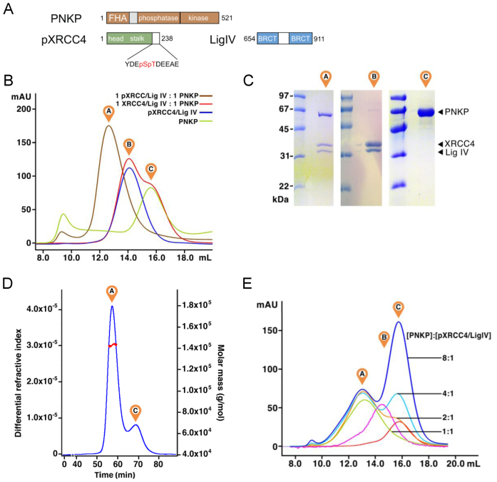
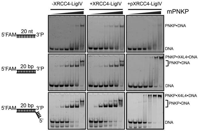
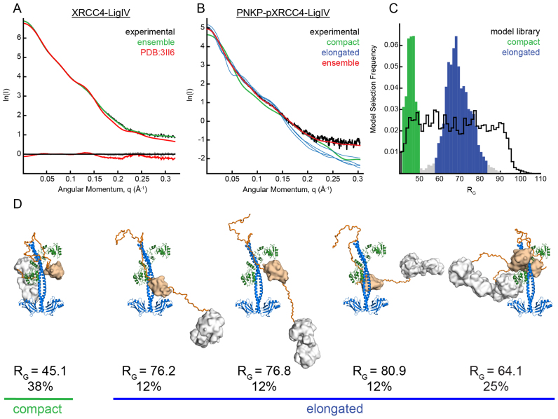
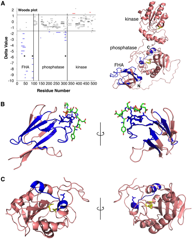
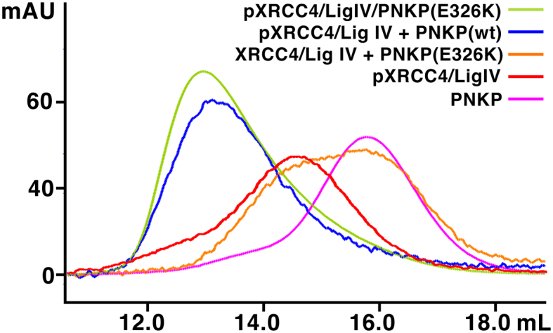
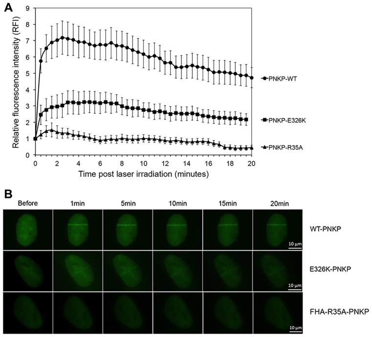
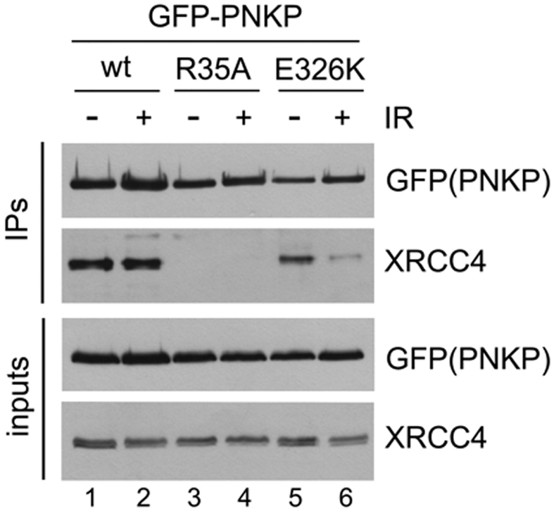
Non-homologous end joining (NHEJ) repairs DNA double strand breaks in non-cycling eukaryotic cells. NHEJ relies on polynucleotide kinase/phosphatase (PNKP), which generates 5΄-phosphate/3΄-hydroxyl DNA termini that are critical for ligation by the NHEJ DNA ligase, LigIV. PNKP and LigIV require the NHEJ scaffolding protein, XRCC4. The PNKP FHA domain binds to the CK2-phosphorylated XRCC4 C-terminal tail, while LigIV uses its tandem BRCT repeats to bind the XRCC4 coiled-coil. Yet, the assembled PNKP-XRCC4-LigIV complex remains uncharacterized. Here, we report purification and characterization of a recombinant PNKP-XRCC4-LigIV complex. We show that the stable binding of PNKP in this complex requires XRCC4 phosphorylation and that only one PNKP protomer binds per XRCC4 dimer. Small angle X-ray scattering (SAXS) reveals a flexible multi-state complex that suggests that both the PNKP FHA and catalytic domains contact the XRCC4 coiled-coil and LigIV BRCT repeats. Hydrogen-deuterium exchange indicates protection of a surface on the PNKP phosphatase domain that may contact XRCC4-LigIV. A mutation on this surface (E326K) causes the hereditary neuro-developmental disorder, MCSZ. This mutation impairs PNKP recruitment to damaged DNA in human cells and provides a possible disease mechanism. Together, this work unveils multipoint contacts between PNKP and XRCC4-LigIV that regulate PNKP recruitment and activity within NHEJ.


Figures

Figure 1.
Figure 1.
Formation of a phosphorylation-dependent 1:2:1 PNKP–pXRCC4–LigIV complex. (A) Overview of the PNKP, XRCC4 and LigIV protein constructs used in this study. (B) Analysis of PNKP interactions with XRCC4–LigIV by Superdex 200 analytical gel filtration chromatography. ‘A’ indicates the elution position of the PNKP–pXRCC4–LigIV complex, ‘B’ indicates the elution position of the XRCC4–LigIV complex, and ‘C’ indicates elution position of free PNKP. (C) Analysis of the peaks in (B) by SDS-PAGE. Molecular weight markers are shown in the left lane for each gel. (D) Analysis of PNKP–pXRCC4–LigIV by MALLS. PNKP–pXRCC4–LigIV, premixed at a 1:2:1 molar ratio was run on a Superose6 gel filtration column with in-line multi-angle laser light scattering (MALLS). The refractive index readout is shown in blue, and the light scattering is shown in red with the eluted peaks labelled as in (B). (E) Titration of PNKP against pXRCC4–LigIV assessed by Superdex 200 analytical gel filtration chromatography as in (B). The numbers refer to the ratio of PNKP:pXRCC4–LigIV for each trace. The magenta trace is a pXRCC4–LigIV control and the red trace is the PNKP control.
Figure 2.
Figure 2.
CK2-phosphorylated XRCC4–LigIV can bind PNKP–DNA substrate complexes. EMSA experiments were carried out using the FAM-labeled DNA constructs illustrated to the left in the absence of XRCC4–LigIV (left panels), with 2 μM XRCC4–LigIV (center panels), or with 2 μM pXRCC4–LigIV (right panels). The PNKP concentrations increased from left to right in each gel. The PNKP concentrations are: 0, 40, 80, 200, 400, 800, 2000 nM.
Figure 3.
Figure 3.
SAXS ensemble analyses indicate PNKP–pXRCC4–LigIV adopts compact and elongated conformations. (A) XRCC41–238/LigIV654–911 experimental X-ray scattering (black) is displayed with the calculated scattering of the XRCC41–203/LigIV654–911 X-ray crystal structure (pdb code 3II6, red), and the scattering calculated from a five model ensemble in which XRCC4 residues 202–238 were allowed to be flexible (green). Residuals are shown below. (B) PNKP–pXRCC4–LigIV experimental X-ray scattering is shown (black) with calculated scattering of a five-model ensemble with χ2 1.7 (red). Four of the theoretical scattering curves for the individual models that constitute the ensemble are shown with curves corresponding to elongated structures in blue, and the single curve corresponding to a compact structure in green. (C) Frequency of occurrence of models extracted in multiple runs of the genetic algorithm plotted against RG. A bi-modal distribution is evident relative to that of the model library (black) indicating a mixture of compact (green) and extended conformations (blue). (D) Models from the ensemble with their RG and the frequency of occurrence. χ2 for fit of individual models to data, from left to right: 13.9; 9.6; 9.5; 15.2; 22.5. Each model is coloured with XRCC4 (blue), Ligase IV (green), polypeptide linkers (orange), PNKP FHA domain (wheat), PNKP catalytic domain (white).
Figure 4.
Figure 4.
HX-MS reveals regions of PNKP FHA and phosphatase domain are protected by pXRCC4–LigIV. (A) Left panel, Woods plot of HX-MS PNKP peptide protection data in which peptides that are significantly protected in complex with pXRCC4–LigIV (Delta value < –1.4) are colored blue while those that show significantly enhanced exchange in the complex (Delta value > 1.4) are colored red. Right panel, overview of the PNKP structure with peptides showing significant protection colored blue. The phosphatase and kinase active sites are colored yellow (PDB ID: 1YJ5). (B) View of protected peptides within the FHA domain. The CK2-phosphorylated peptide from XRCC4 is coloured green (PDB ID: 1YJM). (C) View of the protected peptide within the PNKP phosphatase domain.
Figure 5.
Figure 5.
PNKP E326K is competent to bind pXRCC4–LigIV. One mole equivalent PNKP (wt or E326K) was mixed with one mole equivalent of XRCC4–LigIV complex (+/– phosphorylation) and separated by Superdex 200 analytical gel filtration chromatography as in Figure 1B.
Figure 6.
Figure 6.
E326K mutation abrogates recruitment of PNKP to sites of DNA damage. (A) U2OS cells were transiently transfected with EGFP–PNKP–wt (circles), EGFP–PNKP–R35A (triangles) or EGFP–PNKP–E326K (squares). Laser tracks were introduced after BrdU sensitization and the relative fluorescence determined at 30-s intervals. The means of 30 cells with standard error of the mean at each time point is shown. All data points for EGFP–PNKP–R35A and EGFP–PNKP–E326K were statistically significant (P < 0.05) with respect to EGFP–PNKP–wt. (B) Representative images of laser tracks in cells expressing EGFP–PNKP–wt, EGFP–PNKP–R35A and EGFP–PNKP–E326K. The images show GFP-tagged proteins expressed in the nuclei of individual cells. See Supplementary Figure S5 for an expanded view.
Figure 7.
Figure 7.
E326K mutation reduces the interaction of PNKP with XRCC4. U2OS cells were transfected with EGFP–PNKP–wt, R35A or E326K and after 24 h, either unirradiated (–) or irradiated at 10 Gy and harvested after 1 h. PNKP was immunoprecipitated using anti-GFP antibody and immunoprecipitates were probed with antibodies to GFP (GFP-PNKP) or XRCC4 as shown. The doublets in blots of XRCC4 are likely due to phosphorylation as shown previously (48) and data not shown. The experiment shown is representative of three separate experiments.

References

    1. Radhakrishnan S.K., Jette N., Lees-Miller S.P.. Non-homologous end joining: emerging themes and unanswered questions. DNA Repair (Amst.). 2014; 17:2–8. - PMC - PubMed
    1. Wang C., Lees-Miller S.P.. Detection and repair of ionizing radiation-induced DNA double strand breaks: new developments in nonhomologous end joining. Int. J. Radiat. Oncol. Biol. Phys. 2013; 86:440–449. - PMC - PubMed
    1. Hammel M., Yu Y., Fang S., Lees-Miller S.P., Tainer J.A.. XLF regulates filament architecture of the XRCC4.ligase IV complex. Structure. 2010; 18:1431–1442. - PMC - PubMed
    1. Brouwer I., Sitters G., Candelli A., Heerema S.J., Heller I., de Melo A.J., Zhang H., Normanno D., Modesti M., Peterman E.J. et al. Sliding sleeves of XRCC4-XLF bridge DNA and connect fragments of broken DNA. Nature. 2016; 535:566–569. - PubMed
    1. Grundy G.J., Rulten S.L., Arribas-Bosacoma R., Davidson K., Kozik Z., Oliver A.W., Pearl L.H., Caldecott K.W.. The Ku-binding motif is a conserved module for recruitment and stimulation of non-homologous end-joining proteins. Nat. Commun. 2016; 7:11242. - PMC - PubMed

MeSH terms