. 2017 Aug 1;26(15):2850-2863.
doi: 10.1093/hmg/ddx162.

Progranulin functions as a cathepsin D chaperone to stimulate axonal outgrowth in vivo

Affiliations

Progranulin functions as a cathepsin D chaperone to stimulate axonal outgrowth in vivo

Sander Beel et al. Hum Mol Genet. .

Abstract

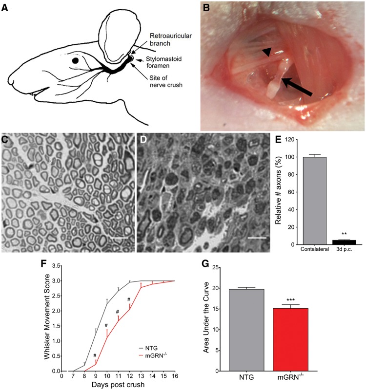
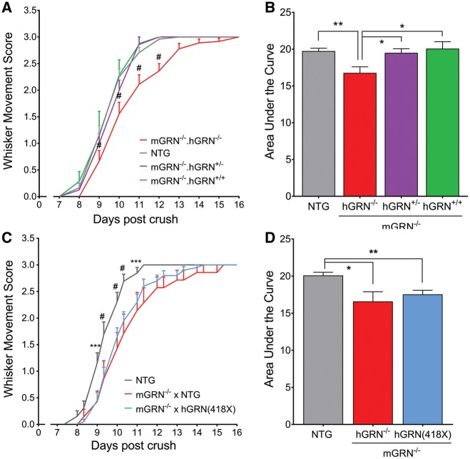
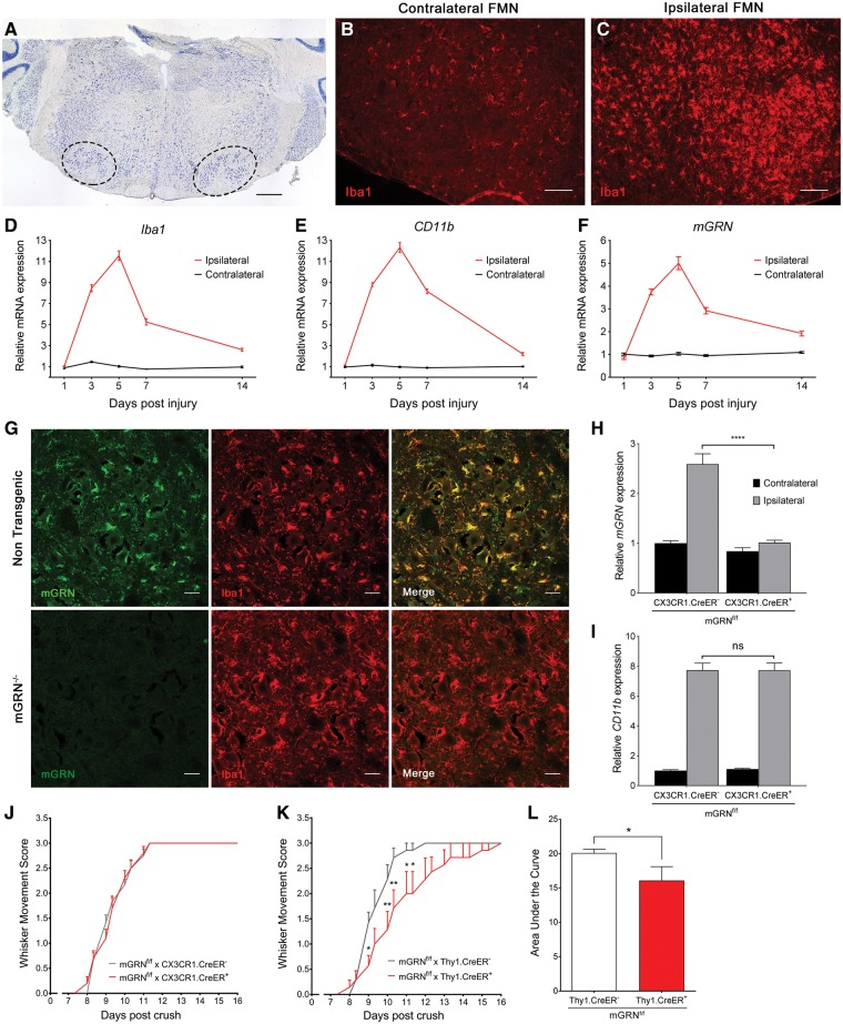
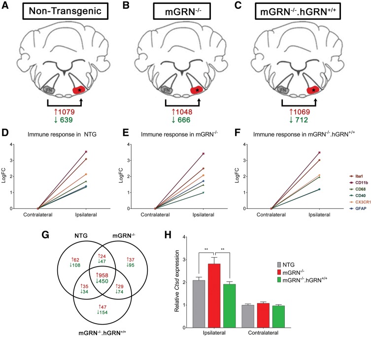
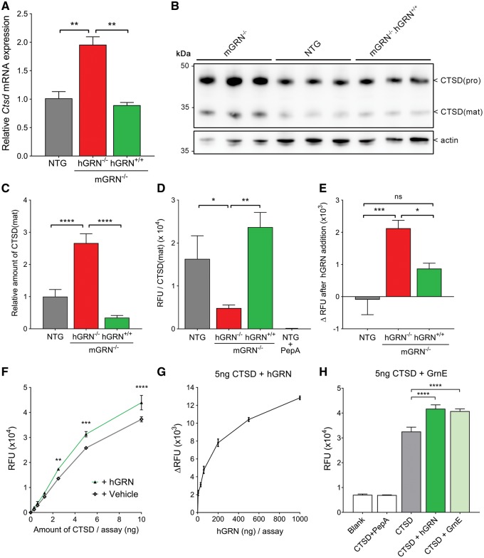
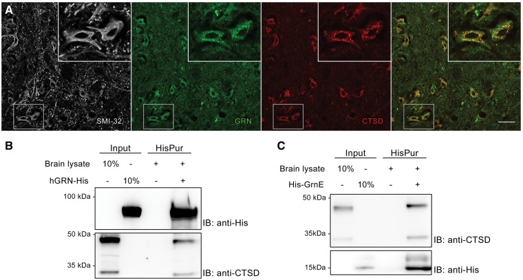
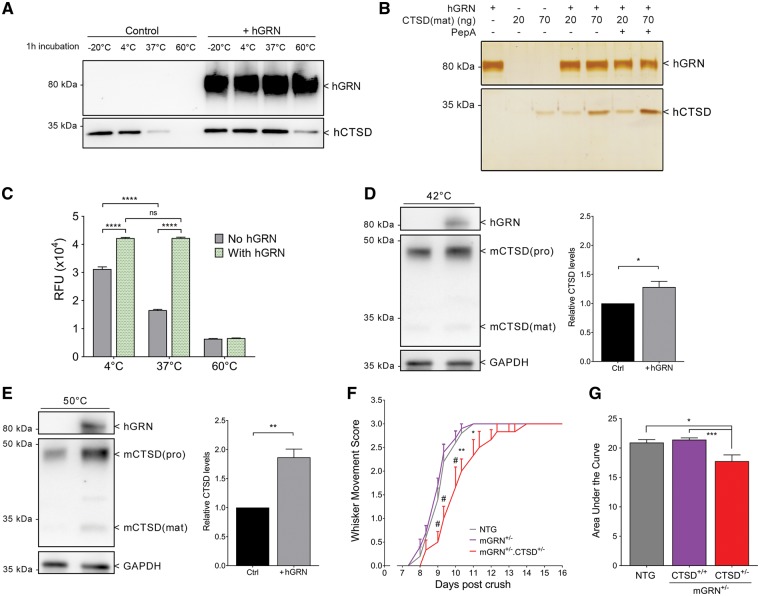
Loss of function mutations in progranulin (GRN) cause frontotemporal dementia, but how GRN haploinsufficiency causes neuronal dysfunction remains unclear. We previously showed that GRN is neurotrophic in vitro. Here, we used an in vivo axonal outgrowth system and observed a delayed recovery in GRN-/- mice after facial nerve injury. This deficit was rescued by reintroduction of human GRN and relied on its C-terminus and on neuronal GRN production. Transcriptome analysis of the facial motor nucleus post injury identified cathepsin D (CTSD) as the most upregulated gene. In aged GRN-/- cortices, CTSD was also upregulated, but the relative CTSD activity was reduced and improved upon exogenous GRN addition. Moreover, GRN and its C-terminal granulin domain granulinE (GrnE) both stimulated the proteolytic activity of CTSD in vitro. Pull-down experiments confirmed a direct interaction between GRN and CTSD. This interaction was also observed with GrnE and stabilized the CTSD enzyme at different temperatures. Investigating the importance of this interaction for axonal regeneration in vivo we found that, although individually tolerated, a combined reduction of GRN and CTSD synergistically reduced axonal outgrowth. Our data links the neurotrophic effect of GRN and GrnE with a lysosomal chaperone function on CTSD to maintain its proteolytic capacity.


Figures

Figure 1
Figure 1
mGRN−/− mice show delayed functional recovery after facial nerve crush injury. (A) Schematic representation of the facial nerve anatomy (adapted and adjusted from Kuzis et al. (55)). The facial nerve crush injury is performed immediately posterior from the bifurcation with the retroauricular branch (B, arrowhead), making the facial nerve completely transparent at the injury site (B, arrow). Semi-thin cross sections of contra- (C) and ipsilateral (D) facial nerve, distal from the injury site. (E) 95% of axons are lost in the distal nerve segment at 3 days post crush. **P < 0.01, Mann-Whitney test. (F) The absence of mGRN significantly delays functional recovery of the of the whisker movement. #P <0.0001, RM Two-way ANOVA followed by Bonferroni correction. (G) Area under the curve of whisker movement recovery scores is significantly reduced in mGRN−/− mice. ***P < 0.001, Student’s t-test. Data is shown as mean ± SEM.
Figure 2
Figure 2
Re-introduction of hGRN rescues whisker movement recovery in mGRN−/− mice, but only if the C-terminal granulin domains are present. (A) mGRN−/− mice that contain one or two copies of the hGRN cDNA show a similar recovery as NTG mice, which is significantly faster than mGRN−/− mice. #P <0.001, RM Two-way ANOVA followed by Bonferroni correction. (B) Analysis of the area under the curve of whisker movement recovery is increased to baseline levels upon introduction of hGRN in mGRN−/− mice. *P <0.05, **P <0.01, One-way ANOVA. (C) The introduction of one or two copies of hGRN(418X) cDNA in mGRN−/− mice did not rescue the delay in whisker movement recovery. ***P <0.001, #P <0.0001, RM Two-way ANOVA followed by Bonferroni correction. (D) Analysis of the are under the curve is still reduced upon introduction of hGRN(418X) in mGRN−/− mice. *P <0.05, **P <0.01, One-way ANOVA. Data is shown as mean ± SEM.
Figure 3
Figure 3
Microgliosis in the facial motor nucleus is maximal at 5 days post injury, but only neuron specific GRN knockout mice recapitulate the delayed recovery of mGRN−/− mice. (A) Thionin stained section through the brainstem. Left and right facial motor nucleus are delineated by the dotted lines. Scale bar= 500 µm. (B–C) A strong increase in Iba1 immunoreactivity is observed in the facial motor nucleus on the ipsilateral side, at 5 days post crush. Scale bar= 100 µm. (D–F) qPCR analysis of microglial genes (Iba1, CD11b) and mGRN, in the facial motor nucleus at different days post crush. (G) mGRN staining overlaps with Iba1 positive microglia and is absent in mGRN−/− mice. Scale bar= 25 µm. (H–I) Relative mRNA expression of mGRN (H) and CD11b (I) in the ipsi- and contralateral facial motor nucleus of microglial specific mGRN knockout mice at 5 days post crush. ****P <0.0001, Two-way ANOVA followed by Tukey’s test. (J–K) Microglial mGRN knockout mice show a normal recovery after crush (J), while neuronal GRN deletion recapitulates the delayed recovery that is observed in mGRN−/− mice (K). *P <0.05, **P <0.01, RM Two-way ANOVA followed by Bonferroni correction. (L) Analysis of area under the curve of whisker movement recovery in Thy1.CreER+ mGRNf/f mice is significantly reduced. *P <0.05, Mann-Whitney test. Data is shown as mean ± SEM. FMN = facial motor nucleus.
Figure 4
Figure 4
Transcriptome analysis of facial motor nucleus RNA, 5 days post injury. (A–C) Schematic representation of the setup used for our RNA sequencing experiment. The number of significantly up- and downregulated genes (FDR< 0.05; LogFC>|1|) post injury is shown in red and green, respectively. (D–F) Graphical representation of the immune response in the three different groups. (G) Venn diagram showing the number of significantly up- and downregulated genes post crush, in red and green, respectively. (H) qPCR validation of CTSD mRNA expression after nerve crush injury confirming a significant upregulation of CTSD in the ipsilateral facial motor nucleus. **P <0.01, Two-way ANOVA followed by Bonferroni correction. Data is shown as mean ± SEM.
Figure 5
Figure 5
GRN and GrnE increase the proteolytic activity of CTSD. (A) CTSD mRNA expression in brains of 22-24 month old mice. **P <0.01, One-way ANOVA. (B) Western blot analysis of old mice brain lysates that were used for CTSD enzymatic assays. (C) Quantification of CTSD(mat) levels, normalized to actin. ****P <0.0001, One-way ANOVA. (D) CTSD enzyme activity in relative fluorescent units (RFU) normalized to the levels of mature CTSD protein. 2.5 µM pepstatin A was added to NTG lysates as a specificity control. *P <0.05, **P <0.01, One-way ANOVA. (E) Change in CTSD enzymatic activity upon the addition of 100 ng recombinant hGRN to brain lysates. *P <0.05, ***P <0.001, One-way ANOVA. (F) Addition of 100 ng hGRN to an active recombinant CTSD peptide increases its proteolytic activity. **P <0.01, ***P <0.001, ****P <0.0001, Two-way ANOVA followed by Bonferroni correction. (G) hGRN increases the proteolytic activity of 5 ng recombinant CTSD in a dose-dependent manner. (H) Addition of equimolar amounts GrnE and GRN increase the proteolytic activity of 5 ng CTSD to a similar extent. ****P <0.0001, One-way ANOVA. Data are shown as mean ± SEM.
Figure 6
Figure 6
GRN and GrnE show a direct interaction with CTSD. (A) GRN and CTSD co-localize inside SMI-32 positive neurons in the facial motor nucleus. Scale bar= 50 µm. (B–C) Pull down of recombinant hGRN (B) or GrnE (C) co-precipitates both the pro- and mature form of CTSD from brain lysates.
Figure 7
Figure 7
GRN stabilizes CTSD, which is necessary to mediate axonal outgrowth in vivo. (A) Heat inactivation of CTSD is prevented by the addition of hGRN. (B) Silver staining showing that the inhibition of CTSD activity by PepA has no effect on its heat inactivation. (C) Pre-incubation of CTSD for 1 h at different temperatures leads to a significant reduction in its proteolytic capacity, which is prevented by the addition of hGRN. ****P < 0.0001, Two-way ANOVA followed by Bonferroni correction. (D,E) Heat inactivation of CTSD in mGRN−/− MEFs for 1 h at 42 °C (D) and 15’ (after 3h pre-incubation with hGRN) at 50 °C (E) is prevented by the supplementation of hGRN, in a temperature dependent manner. *P < 0.05, **P < 0.01, Student’s t-test. (F) A combined heterozygous reduction of mGRN and CTSD significantly delays whisker movement recovery. *P < 0.05, **P < 0.01, #P < 0.0001, RM Two-way ANOVA followed by Bonferroni correction. (G) Analysis of the area under the curve of whisker movement recovery. *P < 0.05, ***P < 0.001, Kruskal Wallis test followed by Dunn’s test. Data is shown as mean ± SEM. PepA = pepstatin A.

References

    1. Bateman A., Bennett H.P. (1998) Granulins: the structure and function of an emerging family of growth factors. J. Endocrinol., 158, 145–151. - PubMed
    1. Zhu J., Nathan C., Jin W., Sim D., Ashcroft G.S., Wahl S.M., Lacomis L., Erdjument-Bromage H., Tempst P., Wright C.D.. et al. (2002) Conversion of proepithelin to epithelins: roles of SLPI and elastase in host defense and wound repair. Cell, 111, 867–878. - PubMed
    1. De Muynck L., Van Damme P. (2011) Cellular effects of progranulin in health and disease. J. Mol. Neurosci., 45, 549–560. - PubMed
    1. Zheng Y., Brady O.A., Meng P.S., Mao Y., Hu F. (2011) C-terminus of progranulin interacts with the beta-propeller region of sortilin to regulate progranulin trafficking. PLoS One, 6, e21023.. - PMC - PubMed
    1. Petkau T.L., Leavitt B.R. (2014) Progranulin in neurodegenerative disease. Trends Neurosci., 37, 388–398. - PubMed