

Validation of biomechanical deformable image registration in the abdomen, thorax, and pelvis in a commercial radiotherapy treatment planning system
- PMID: 28453911
- PMCID: PMC5535790
- DOI: 10.1002/mp.12307
Validation of biomechanical deformable image registration in the abdomen, thorax, and pelvis in a commercial radiotherapy treatment planning system
Abstract
Purpose: The accuracy of deformable image registration tools can vary widely between imaging modalities and specific implementations of the same algorithms. A biomechanical model-based algorithm initially developed in-house at an academic institution was translated into a commercial radiotherapy treatment planning system and validated for multiple imaging modalities and anatomic sites.
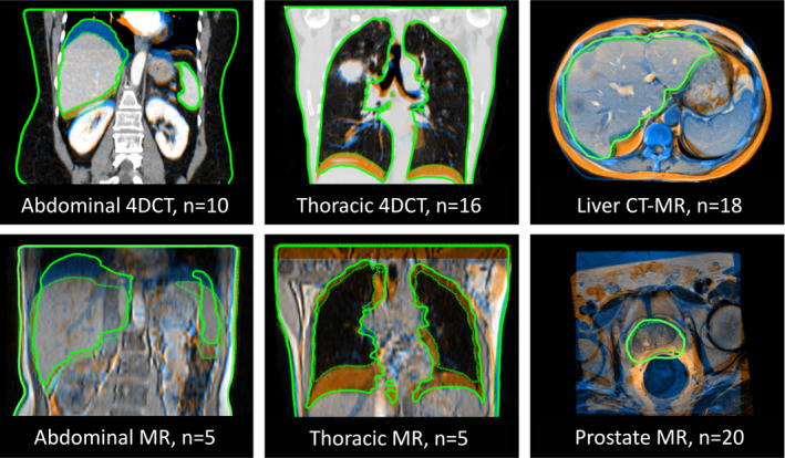
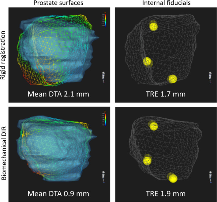
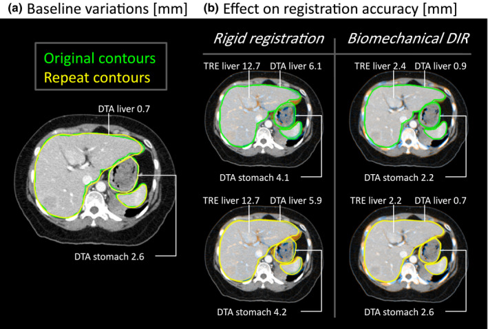
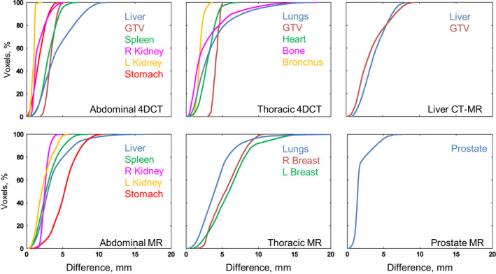
Methods: Biomechanical deformable registration (Morfeus) is a geometry-driven algorithm based on the finite element method. Boundary conditions are derived from the model-based segmentation of controlling structures in each image which establishes a point-to-point surface correspondence. For each controlling structure, material properties and fixed or sliding interfaces are assigned. The displacements of internal volumes for controlling structures and other structures implicitly deformed are solved with finite element analysis. Registration was performed for 74 patients with images (mean vector resolution) of thoracic and abdominal 4DCT (2.8 mm) and MR (5.3 mm), liver CT-MR (4.5 mm), and prostate MR (2.6 mm). Accuracy was quantified between deformed and actual target images using distance-to-agreement (DTA) for structure surfaces and the target registration error (TRE) for internal point landmarks.
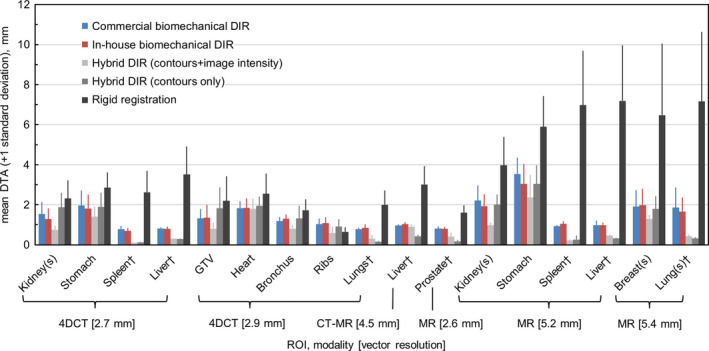
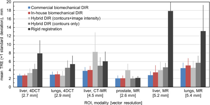
Results: The results of the commercial implementation were as follows. The mean DTA was ≤ 1.0 mm for controlling structures and 1.0-3.5 mm for implicitly deformed structures on average. TRE ranged from 2.0 mm on prostate MR to 5.1 mm on lung MR on average, within 0.1 mm or lower than the image voxel sizes. Accuracy was not overly sensitive to changes in the material properties or variability in structure segmentations, as changing these inputs affected DTA and TRE by ≤ 0.8 mm. Maximum DTA > 5 mm occurred for 88% of the structures evaluated although these were within the inherent segmentation uncertainty for 82% of structures. Differences in accuracy between the commercial and in-house research implementations were ≤ 0.5 mm for mean DTA and ≤ 0.7 mm for mean TRE.
Conclusions: Accuracy of biomechanical deformable registration evaluated on a large cohort of images in the thorax, abdomen and prostate was similar to the image voxel resolution on average across multiple modalities. Validation of this treatment planning system implementation supports biomechanical deformable registration as a versatile clinical tool to enable accurate target delineation at planning and treatment adaptation.
Keywords: biomechanical models; deformable image registration; multimodality imaging.
© 2017 American Association of Physicists in Medicine.
Conflict of interest statement
K.K. Brock, J.L. Moseley, and D.A. Jaffray have a licensing agreement with RaySearch Laboratories for the deformable registration technology in this study. S. Svensson and B. Hårdemark are employees of RaySearch Laboratories.
Figures
References
-
- Kumarasiri A, Siddiqui F, Liu C, et al. Deformable image registration based automatic CT‐to‐CT contour propagation for head and neck adaptive radiotherapy in the routine clinical setting. Med Phys. 2014;41:121712. - PubMed
-
- Speight R, Sykes J, Lindsay R, Franks K, Thwaites D. The evaluation of a deformable image registration segmentation technique for semi‐automating internal target volume (ITV) production from 4DCT images of lung stereotactic body radiotherapy (SBRT) patients. Radiother Oncol. 2011;98:277–283. - PubMed
-
- Swaminath A, Massey C, Brierley JD, et al. Accumulated delivered dose response of stereotactic body radiation therapy for liver metastases. Int J Radiat Oncol Biol Phys. 2015;93:639–648. - PubMed
-
- Brock KK, Velec M, Lee JHM. Validation of image registration. In: Brock KK, ed. Image Processing in Radiation Therapy. Boca Raton: CRC Press; 2013:41–62.
MeSH terms
Grants and funding
LinkOut - more resources
Full Text Sources
Other Literature Sources
Medical
