

Expression and prognostic relevance of MAGE-A3 and MAGE-C2 in non-small cell lung cancer
- PMID: 28454298
- PMCID: PMC5403542
- DOI: 10.3892/ol.2017.5665
Expression and prognostic relevance of MAGE-A3 and MAGE-C2 in non-small cell lung cancer
Abstract
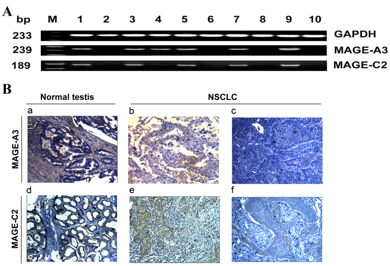
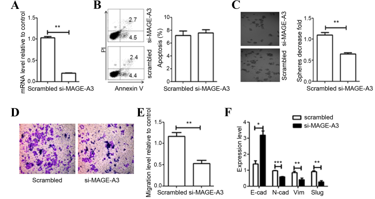
Melanoma-associated antigen (MAGE)-A3 and MAGE-C2 are antigens encoded by cancer-germline genes, and have been recognized as potential prognostic biomarkers and attractive targets for immunotherapy in multiple types of cancer. The present study aimed to analyze the clinicopathological significance of MAGE-A3/C2 expression in non-small cell lung cancer (NSCLC). The association between MAGE-A3/C2 mRNA and protein expression, and the pathological characteristics and overall survival of patients with NSCLC was analyzed. In addition, the functional role of MAGE-A3 in human NSCLC cell line A549 was examined in vitro. MAGE-A3/C2 mRNA expression was identified in 73% (151/206) and 53% (109/206) of patients with NSCLC, respectively. MAGE-A3/C2 protein expression was identified in 58% (44/76) and 53% (40/76) of NSCLC cases, respectively. MAGE-A3 mRNA expression was observed to be associated with smoking history, disease stage and lymph node metastasis. However, no association was identified between MAGE-C2 mRNA expression and the clinicopathological characteristics of patients with NSCLC. MAGE-A3/C2-positive patients had a poorer survival rate compared with MAGE-A3/C2-negative patients. Multivariate analysis identified that MAGE-A3 expression may serve as an independent marker of poor prognosis in patients with NSCLC. Downregulation of MAGE-A3 mRNA expression in A549 cells resulted in lower migration and colony formation rates, and a higher amount of epithelial marker and lower amount of mesenchymal marker expression compared with the control group. These results indicate that MAGE-A3 serves a role in NSCLC cell metastasis through the induction of epithelial-mesenchymal transition. In conclusion, MAGE-A3 may serve as a diagnostic and prognostic biomarker for patients with NSCLC, due to its association with tumor progression and poor clinical outcome.
Keywords: cancer-germline genes; epithelial-mesenchymal transition; non-small cell lung cancer; prognostic biomarker.
Figures

References
-
- Quoix E, Lena H, Losonczy G, Forget F, Chouaid C, Papai Z, Gervais R, Ottensmeier C, Szczesna A, Kazarnowicz A. TG4010 immunotherapy and first-line chemotherapy for advanced non-small-cell lung cancer (TIME): Results from the phase 2b part of a randomised, double-blind, placebo-controlled, phase 2b/3 trial. Lancet Oncol. 2016;17:212–223. doi: 10.1016/S1470-2045(15)00483-0. - DOI - PubMed
-
- Pinato DJ, Shiner RJ, White SD, Black JR, Trivedi P, Stebbing J, Sharma R, Mauri FA. Intra-tumoral heterogeneity in the expression of programmed-death (PD) ligands in isogeneic primary and metastatic lung cancer: Implications for immunotherapy. Oncoimmunology. 2016;5:e1213934. doi: 10.1080/2162402X.2016.1213934. - DOI - PMC - PubMed
LinkOut - more resources
Full Text Sources
Other Literature Sources
Miscellaneous