. 2017 Apr;13(4):2034-2044.
doi: 10.3892/ol.2017.5697. Epub 2017 Feb 8.

Modeling nasopharyngeal carcinoma in three dimensions

Affiliations

Modeling nasopharyngeal carcinoma in three dimensions

Prabu Siva Sankar et al. Oncol Lett. 2017 Apr.

Abstract

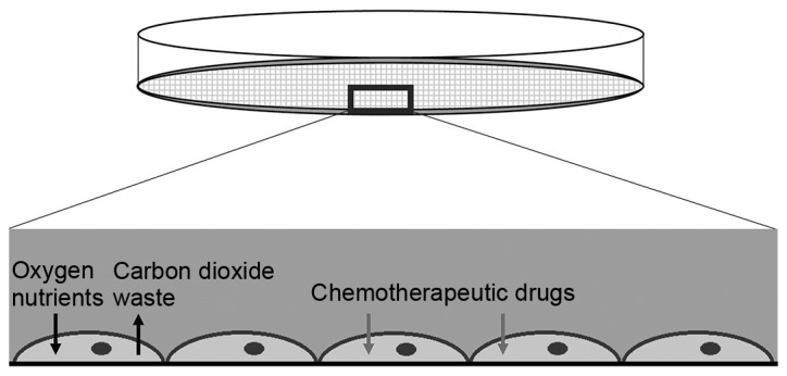
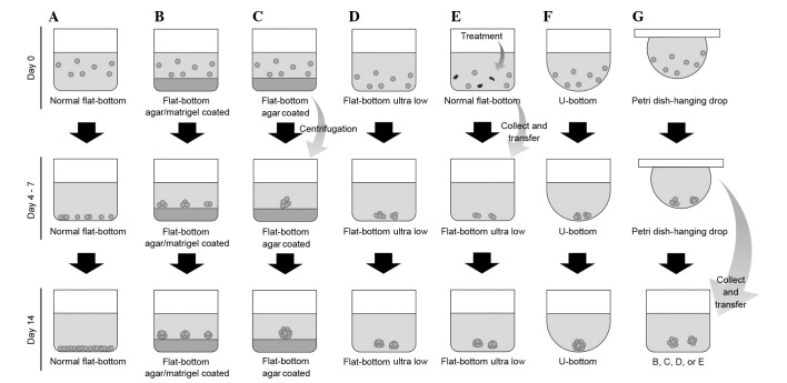
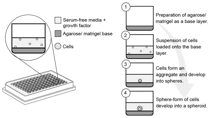
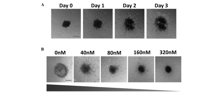
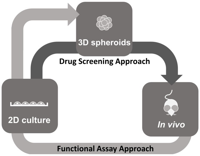
Nasopharyngeal carcinoma (NPC) is a type of cancer endemic in Asia, including Malaysia, Southern China, Hong Kong and Taiwan. Treatment resistance, particularly in recurring cases, remains a challenge. Thus, studies to develop novel therapeutic agents are important. Potential therapeutic compounds may be effectively examined using two-dimensional (2D) cell culture models, three-dimensional (3D) spheroid models or in vivo animal models. The majority of drug assessments for cancers, including for NPC, are currently performed with 2D cell culture models. This model offers economical and high-throughput screening advantages. However, 2D cell culture models cannot recapitulate the architecture and the microenvironment of a tumor. In vivo models may recapitulate certain architectural and microenvironmental conditions of a tumor, however, these are not feasible for the screening of large numbers of compounds. By contrast, 3D spheroid models may be able to recapitulate a physiological microenvironment not observed in 2D cell culture models, in addition to avoiding the impediments of in vivo animal models. Thus, the 3D spheroid model offers a more representative model for the study of NPC growth, invasion and drug response, which may be cost-effective without forgoing quality.

Keywords: 2-dimensional cell culture model; 3-dimensional spheroid model; in vivo models; nasopharyngeal carcinoma; spheroids.


Figures

Figure 1.
Figure 1.
2D cell culture model. Cells grown in 2D cell culture adhere to the plastic surface of the plate. They receive maximum exposure to drugs with optimal diffusion of nutrients and waste products. The 2D culture model provides the cells with the optimal conditions to proliferate, thus, inducing high proliferative rates, although this does not reflect the tumor behavior in vivo. 2D, two-dimensional.
Figure 2.
Figure 2.
Similarities between the 3D spheroid model and a tumor in vivo. The spheroid received nutrients from the medium and the tumor through its vascularization, although they are subject to concentration gradients, which limit efficient diffusion. This creates a microenvironment, leading to various conditions of cells within the tumors and spheroids. The necrotic core exists due to the lack of available nutrients and the accumulation of waste, whereas the increased metabolic activity at the periphery is due to the more efficient diffusion of nutrients and waste. In each condition, the cells exist in a 3D conformation with interactions with the extracellular matrix. 3D, three-dimensional.
Figure 3.
Figure 3.
Comparison between various methods and plate types used for the generation of 3D spheroids. (A) Normal flat-bottom cell culture plate; (B) Normal flat-bottom cell culture plate coated with agarose or Matrigel; (C) Normal flat-bottom cell culture plate coated with agarose; the cells clump due to centrifugation; (D) Ultra-Low attachment plate; (E) Modification of drug resistant spheroid: drug treatment is conducted in a ultra-low attachment plate prior to spheroid culture; (F) U-bottom plate; (G) Hanging drop method using a petri dish: following the formation of cell clumps, the aggregates may be transferred into plate B, C, D or E. 3D, three-dimensional.
Figure 4.
Figure 4.
Formation of spheroids using either agarose or Matrigel as the base layer. Various concentrations of agarose or Matrigel may be applied as the base layer. The cells are suspended in serum-free medium with addition of growth factors such as basic fibroblast growth factor and epidermal growth factor. After four days, cells aggregate and form spheroids.
Figure 5.
Figure 5.
Growth and invasion of the HK1 spheroids into the collagen matrix over three days. (A) Phase contrast images of spheroid growth and invasion were taken every 24 h using the Nikon Ti Eclipse inverted fluorescence microscope; scale bar, 200 µm (B) Spheroids were treated with the chemotherapeutic drug Flavopiridol at the concentrations indicated. Phase contrast images taken every 24 h reveal that Flavopiridol induced a dose-dependent inhibition of spheroid growth and invasion (slope represents level of growth and invasion of the spheroids); scale bar, 200 µm.
Figure 6.
Figure 6.
Proposed application of 3D spheroid models for therapeutic studies. Drug Screening Approach (dark grey arrow): The traditional 2D model is more economical and suitable for high-throughput screening compared with the 3D model. However, 2D models do not represent human cancer as in vivo models do. The transition from the 2D to the 3D model may allow for a relatively high throughput screening with the advantage of a more physiological model. The 3D model mimics the complex in vivo microenvironment, particularly with regard to gene expression, signaling pathways and drug sensitivity. The transition from the 3D model to animal models may also reduce the number of animals used for drug testing and may be more economical as animal models are costly and time-consuming. By using 3D models, more detailed data regarding drug suitability may be obtained prior to initiating in vivo studies with animal models. Functional Assay Approach (light grey arrow): In vivo tumor models may also be developed as in vitro 3D spheroid models for high-throughput functional assays. Cell lines can first be established and characterized in 2D from tumors obtained from the in vivo models such as the genetically engineered mouse models and xenografts and transitioned into 3D spheroids for functional studies. 2D, two-dimensional; 3D, three-dimensional.

References

    1. Loh LE, Chee TS, John AB. The anatomy of the Fossa of Rosenmuller-its possible influence on the detection of occult nasopharyngeal carcinoma. Singapore Med J. 1991;32:154–155. - PubMed
    1. Pua KC, Khoo AS, Yap YY, Subramaniam SK, Ong CA, Krishnan G Gopala, Shahid H. Malaysian Nasopharyngeal Carcinoma Study Group: Nasopharyngeal Carcinoma Database. Med J Malaysia. 2008;63:59–62. (Suppl C) - PubMed
    1. Khoo ASB, Pua KC. In: Nasopharyngeal Carcinoma: Keys for Translational Medicine and Biology. Busson P, editor. Landes Bioscience; Austin, TX: 2013. - DOI
    1. Lo KW, Chung GT, To KF. Deciphering the molecular genetic basis of NPC through molecular, cytogenetic, and epigenetic approaches. Semin Cancer Biol. 2012;22:79–86. doi: 10.1016/j.semcancer.2011.12.011. - DOI - PubMed
    1. Chan AT, Hsu MM, Goh BC, Hui EP, Liu TW, Millward MJ, Hong RL, Whang-Peng J, Ma BB, To KF, et al. Multicenter, phase II study of cetuximab in combination with carboplatin in patients with recurrent or metastatic nasopharyngeal carcinoma. J Clin Oncol. 2005;23:3568–3576. doi: 10.1200/JCO.2005.02.147. - DOI - PubMed