. 2017 Jul 1;313(1):H125-H137.
doi: 10.1152/ajpheart.00842.2016. Epub 2017 Apr 28.

Quantitative cardiac phosphoproteomics profiling during ischemia-reperfusion in an immature swine model

Affiliations

Quantitative cardiac phosphoproteomics profiling during ischemia-reperfusion in an immature swine model

Dolena Ledee et al. Am J Physiol Heart Circ Physiol. .

Abstract

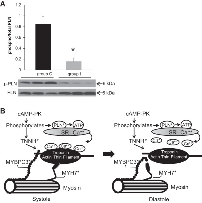
Ischemia-reperfusion (I/R) results in altered metabolic and molecular responses, and phosphorylation is one of the most noted regulatory mechanisms mediating signaling mechanisms during physiological stresses. To expand our knowledge of the potential phosphoproteomic changes in the myocardium during I/R, we used Isobaric Tags for Relative and Absolute Quantitation-based analyses in left ventricular samples obtained from porcine hearts under control or I/R conditions. The data are available via ProteomeXchange with identifier PXD006066. We identified 1,896 phosphopeptides within left ventricular control and I/R porcine samples. Significant differential phosphorylation between control and I/R groups was discovered in 111 phosphopeptides from 86 proteins. Analysis of the phosphopeptides using Motif-x identified five motifs: (..R..S..), (..SP..), (..S.S..), (..S…S..), and (..S.T..). Semiquantitative immunoblots confirmed site location and directional changes in phosphorylation for phospholamban and pyruvate dehydrogenase E1, two proteins known to be altered by I/R and identified by this study. Novel phosphorylation sites associated with I/R were also identified. Functional characterization of the phosphopeptides identified by our methodology could expand our understanding of the signaling mechanisms involved during I/R damage in the heart as well as identify new areas to target therapeutic strategies.NEW & NOTEWORTHY We used Isobaric Tags for Relative and Absolute Quantitation technology to investigate the phosphoproteomic changes that occur in cardiac tissue under ischemia-reperfusion conditions. The results of this study provide an extensive catalog of phosphoproteins, both predicted and novel, associated with ischemia-reperfusion, thereby identifying new pathways for investigation.

Keywords: Isobaric Tags for Relative and Absolute Quantitation; ischemia-reperfusion; phosphoproteomics.


Figures

Fig. 1.
Fig. 1.
Phosphoproteome analysis of cardiac tissue after ischemia-reperfusion (I/R). Schema illustrates the workflow of control or I/R-treated piglets. The total number of phosphopeptides are identified, and the distributions of the identified sites are according to the serine (s*), threonine (t*), and tyrosine (y*) amino acid are shown. iTRAQ, Isobaric Tags for Relative and Absolute Quantitation; LC MS/MS, liquid chromatography-tandem mass spectroscopy.
Fig. 2.
Fig. 2.
Cluster analysis of the differentially expressed phosphoproteins. Each row represents a single protein, and each column represents a single sample. The data are colored red for decreased phosphorylation and green for increased phosphorylation. Gene names designate the protein (n = 4 per group).
Fig. 3.
Fig. 3.
A: protein-protein interaction network among phosphoproteins. The line thickness represents the supported confidence (correlation coefficient) between nodes. Nodes with red and black borders are phosphoproteins with increased and decreased phosphorylated sites in the I/R group compared with the control group, respectively. The green-lettered phosphoproteins contain multiple phosphopeptide sites, and the phosphoproteins with green borders have multiple phosphorylation sites with both increased and decreased phosphorylation states. The grayed-in phosphoproteins were confirmed by immunoblot analysis. The majority of the phosphoproteins (unencapsulated) have roles in cell junctions. The phosphoproteins encircled in purple have roles in RNA processing, and the phosphoproteins encircled in blue have roles in energy production. B: 39 of the 86 altered phosphoproteins had no associative network identified based on this data set.
Fig. 4.
Fig. 4.
A: representative blots for expression profiles of phospholamban (PLN) and phosphorylated (p-)PLN (Ser16) comparing control and I/R treatment. Phosphorylation levels were normalized to total levels. Values are means ± SE; n = 3 per group. All experiments were performed in duplicate. *P < 0.01. B: illustration of the cardiac muscle contraction pathway. *Highlights phosphoproteins identified in this study as significantly (de)phosphorylated. TNNI1, troponin I; MYBPC3, myosin-binding protein-C; MYH7, myosin 7.
Fig. 5.
Fig. 5.
A: representative blots for expression profiles of total pyruvate dehydrogenase (PDH) and p-PDH (Ser293) comparing control and I/R treatment. Phosphorylation levels were normalized to total levels. Values are means ± SE; n = 3 per group. All experiments were performed in duplicate. *P < 0.01. B: illustration of acetyl-CoA synthesis pathways. *Highlights phosphopeptides identified in this study as significantly (de)phosphorylated. DLAT, dihydrolipoamide S-acetyltransferase; DLD, dihydrolipoamide dehydrogenase; PDHX, E3-binding protein; ACSS2, acetyl-CoA synthetase, PDK, pyruvate dehydrogenase kinases; PDP, PDH phosphatase; DCA, dichloroacetate.

References

    1. Abdul-Ghani S, Heesom KJ, Angelini GD, Suleiman MS. Cardiac phosphoproteomics during remote ischemic preconditioning: a role for the sarcomeric Z-disk proteins. BioMed Res Int 2014: 767812, 2014. doi:10.1155/2014/767812. - DOI - PMC - PubMed
    1. Alessi DR, Caudwell FB, Andjelkovic M, Hemmings BA, Cohen P. Molecular basis for the substrate specificity of protein kinase B; comparison with MAPKAP kinase-1 and p70 S6 kinase. FEBS Lett 399: 333–338, 1996. doi:10.1016/S0014-5793(96)01370-1. - DOI - PubMed
    1. Ambrus A, Torocsik B, Tretter L, Ozohanics O, Adam-Vizi V. Stimulation of reactive oxygen species generation by disease-causing mutations of lipoamide dehydrogenase. Hum Mol Genet 20: 2984–2995, 2011. doi:10.1093/hmg/ddr202. - DOI - PubMed
    1. Ambrus A, Wang J, Mizsei R, Zambo Z, Torocsik B, Jordan F, Adam-Vizi V. Structural alterations induced by ten disease-causing mutations of human dihydrolipoamide dehydrogenase analyzed by hydrogen/deuterium-exchange mass spectrometry: Implications for the structural basis of E3 deficiency. Biochim Biophys Acta 1862: 2098–2109, 2016. doi:10.1016/j.bbadis.2016.08.013. - DOI - PMC - PubMed
    1. Bagheri F, Khori V, Alizadeh AM, Khalighfard S, Khodayari S, Khodayari H. Reactive oxygen species-mediated cardiac-reperfusion injury: Mechanisms and therapies. Life Sci 165: 43–55, 2016. doi:10.1016/j.lfs.2016.09.013. - DOI - PubMed