. 2017 Aug;23(8):1209-1223.
doi: 10.1261/rna.060467.116. Epub 2017 Apr 28.

Impact of RNA structure on ZFP36L2 interaction with luteinizing hormone receptor mRNA

Affiliations

Impact of RNA structure on ZFP36L2 interaction with luteinizing hormone receptor mRNA

Christopher B Ball et al. RNA. 2017 Aug.

Abstract

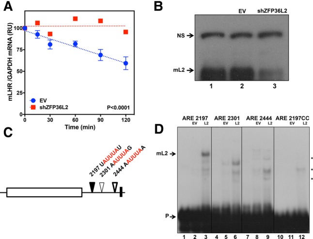
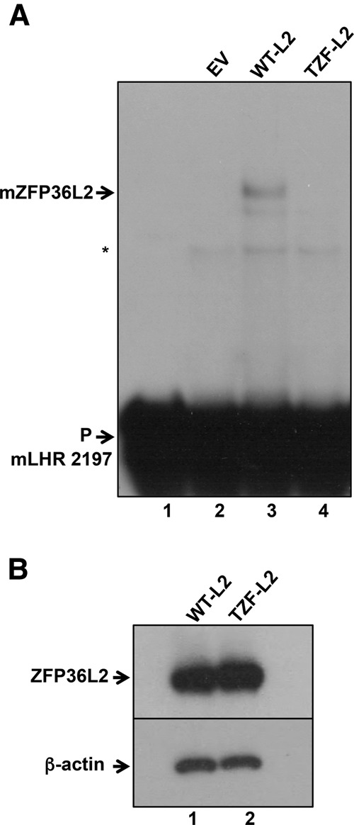
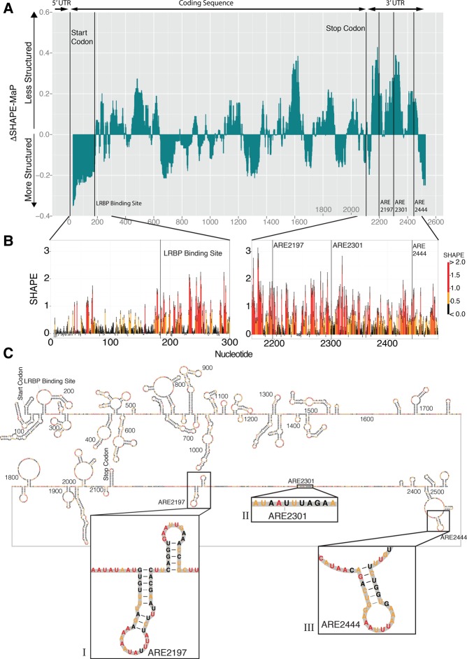
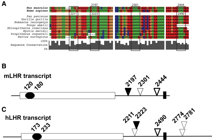
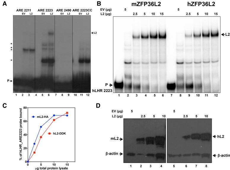
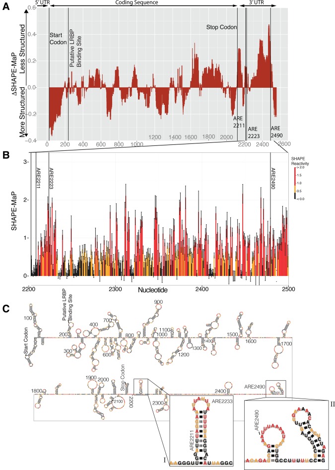
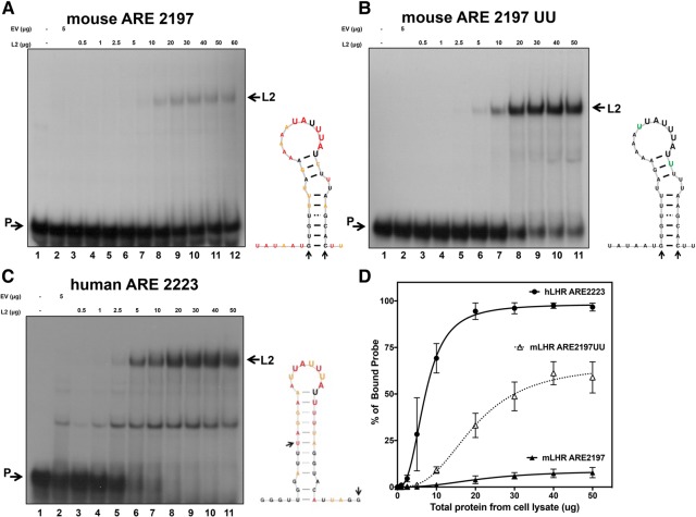
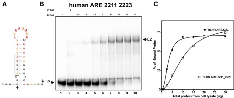
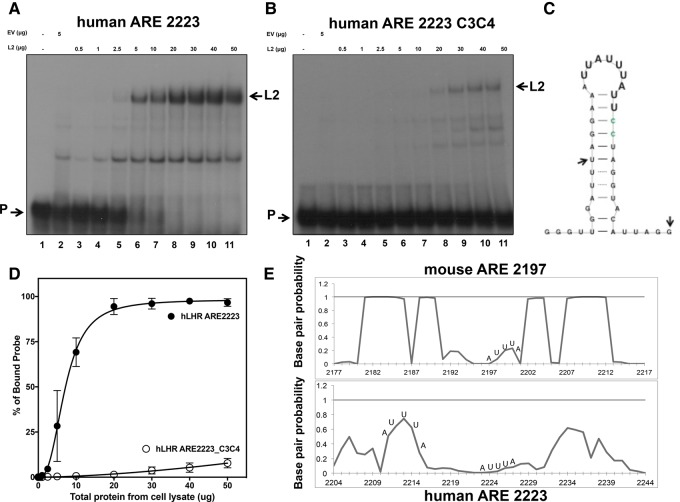
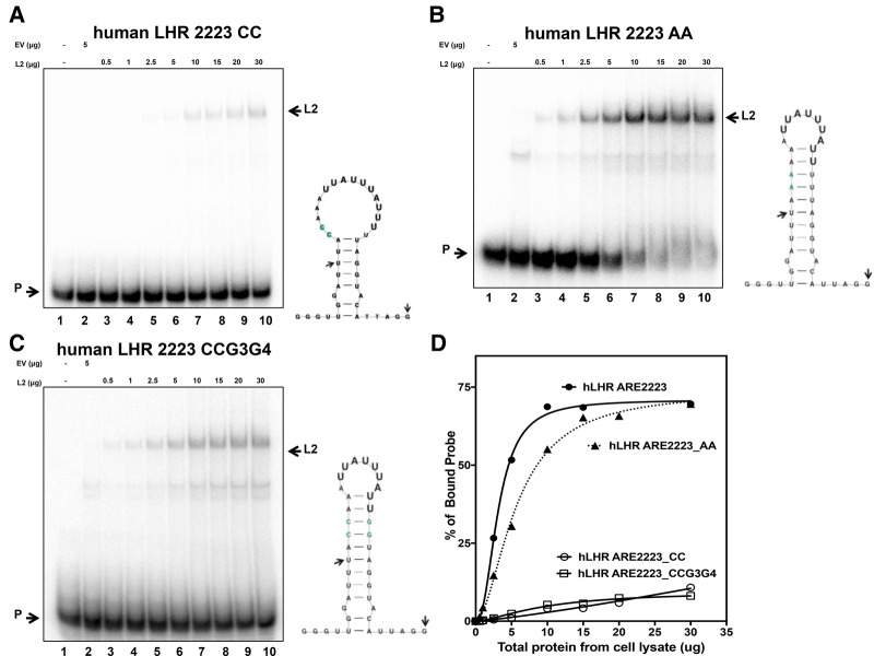
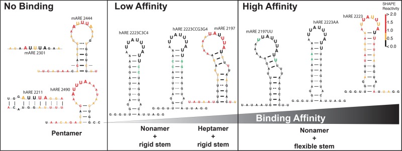
ZFP36L2 (L2) destabilizes AU-rich element (ARE)-containing transcripts and has been implicated in female fertility. We have shown that only one of three putative AREs within the 3' UTR of murine luteinizing hormone receptor mRNA, ARE2197 (UAUUUAU), is capable of interacting with L2. To assess whether structural elements of ARE2197 could explain this unique binding ability, we performed whole-transcript SHAPE-MaP (selective 2' hydroxyl acylation by primer extension-mutational profiling) of the full-length mLHR mRNA. The data revealed that the functional ARE2197 is located in a hairpin loop structure and most nucleotides are highly reactive. In contrast, each of the nonbinding AREs, 2301 and 2444, contains only a pentamer AUUUA; and in ARE2301 much of the ARE sequence is poorly accessible. Because the functional mARE was also found to be conserved in humans at the sequence level (ARE 2223), we decided to investigate whether binding and structure are also preserved. Similar to mouse, only one ARE in hLHR mRNA is capable of binding to L2; and it is also located in a hairpin structure, based on our SHAPE-MaP data. To investigate the role of secondary structure in the binding, we mutated specific nucleotides in both functional AREs. Mutations in the flexible stem region proximal to the loop that enforce strong base-pairing, drastically reduced L2 binding affinity; this confirms that the structural context is critical for L2 recognition of hARE2223. Collectively, our results suggest that a combination of minimal ARE sequence, placement of the ARE in a hairpin loop, and stem flexibility mediate high-affinity L2 binding to hLHR mRNA.

Keywords: LHR mRNA; RNA-binding protein; ZFP36L2; infertility; post-transcriptional modulation.


Figures

FIGURE 1.
FIGURE 1.
mLHR mRNA decay is dependent on ZFP36L2. (A) MLTC1 cells infected with lentivirus without (EV) or with an shRNA targeting ZFP36L2 (LV9) were treated with actinomycin D (4 µg/mL) for 30 min before the time course initiation. Total RNA was harvested from cells at six different time points. LHR and GAPDH mRNAs were quantitated in triplicate by qRT-PCR. The graphic was built from results of two biological replicates. The slopes are different (P < 0.0001). (B) Immunoblot using protein extracts confirmed knockdown of ZFP36L2 (lane 3) in comparison to WT MLTC-1 cells (lane 1) or cells treated with the empty vector (lane 2). (NS) Nonspecific, cross-reacting protein serving as loading control. (C) Schematic location of AREs present in the mouse LHR transcript. AREs located at the 3′ UTR are represented by triangles followed by its respective number, each pentamer sequence composing the ARE (red) is flanked by one nucleotide. (D) RNA electrophoretic mobility shift assays were performed by incubating protein extracts from HEK 293 cells transfected with either the empty vector (EV, 5 µg, lanes 2,5,8,11) or with a vector expressing mZFP36L2-HA (L2, 10 µg, lanes 3,6,9,12) with 0.2 × 10⁵ cpm of mLHR ARE probes. In probe 2197CC, both “As” of ARE2197 were mutated to “Cs.” Lanes 1,4,7,10 contain each probe without any protein extracts, but only buffer. When 5 µg of protein extracts lacking ZFP36L2 protein (lanes 2,5,8,11) were incubated with each probe, faint bands appeared (indicated by asterisks). They correspond to complexes of endogenous proteins present in HEK 293 cells that interact with the probes. These bands are more evident in lanes 3,6,9,12, because more total protein extracts (10 µg) containing overexpressed ZFP36L2 were used.
FIGURE 2.
FIGURE 2.
C176S mutation in mZFP36L2 abolishes binding to the mLHR ARE 2197 probe. (A) RNA electrophoretic mobility shift assays were performed by incubating protein extracts from HEK 293 cells transfected with the empty vector (EV, 5 µg, lane 2), a vector expressing mZFP36L2-HA (WT-L2) or TZF-Mut-mZFP36L2(C176S)-HA (TZF-L2) (10 µg of each in lanes 3 and 4, respectively), with 0.2 × 10⁵ cpm of the mLHR ARE 2197 probe. The input probe in buffer was analyzed in lane 1. (B) Immunoblotting of 10 µg of protein extracts from HEK 293 cells transfected with mZFP36L2-HA (WT-L2, lane 1), or TZF-Mut-mZFP36L2-HA (TZF-L2, lane 2), were probed with an anti-HA antibody (upper panel). An anti-β actin antibody was used as a loading control (lower panel). These protein extracts were aliquots of the same material used in lanes 3 and 4 of the gel shift assay shown in panel A, respectively.
FIGURE 3.
FIGURE 3.
Structural architecture of mouse LHR mRNA. (A) In vitro 1M7 reactivity for the full-length mouse LHR transcript was obtained and represented as median reactivity relative to the global median. The median-averaged signal was used to identify regions of high-median SHAPE reactivity (above zero), which correspond to less structured areas. Similarly, SHAPE nucleotides with a low median reactivity (below zero) correspond to more structured areas in the transcript. (B) SHAPE data for each individual nucleotide composing the 5′ end of LHR transcript (left) and 3′ UTR illustrate a difference between high structure (low SHAPE) and low structure (high SHAPE). In general, the median SHAPE reactivity increases from 5′ to 3′, indicating less base-pairing in the 3′ UTR. (C) SHAPE-informed secondary structure model of mouse LHR transcript. Zooming in the functional ARE 2197 reveals a typical hairpin (inset I). AREs 2301 and 2444 are shown in insets II and III, respectively.
FIGURE 4.
FIGURE 4.
Multispecies alignment of Adenine–Uridine-rich elements (AREs) within the 3′ untranslated region of luteinizing hormone receptor transcript. (A) Conserved AREs are outlined by black boxes. Mouse (Mus musculus) and Human (Homo sapiens) AREs are numbered according to the position of the first 5′ adenosine of the ARE. Conservation was computed for each position in the alignment and reported as gray vertical bars below each column. Mouse ARE 2197 and 2444 are highly conserved, while human ARE 2211 is present only in some primate species. Schematic drawing of mouse (B) and human (C) LHR transcripts. The lines represent untranslated regions, and the open rectangles correspond to coding sequence. The relative location of each ARE is represented by a triangle. The black rectangle at the 3′ UTR illustrates the location of the poly(A) signal. Two of these three AREs are present in mouse and human (bold and black triangles). However, only ARE 2197 binds ZFP36L2 (black triangles), whereas the conserved ARE 2444 did not show binding (bold triangles). mARE 2301 and hARE 2211 are not conserved (plain triangles). Downstream from the poly(A) signal, the human LHR transcript has two other AREs (2774 and 2781) that are not present in the mouse mRNA, and they were not investigated here. Within the coding region of the mouse and human transcripts, a polypyrimidine rich track corresponds to the LRBP binding site (dark ellipse).
FIGURE 5.
FIGURE 5.
mZFP36L2 and hZFP36L2 bind to ARE2223 from the human LHR transcript. (A) RNA electrophoretic mobility shift assays were performed by incubating protein extracts from HEK 293 cells transfected with empty vector (EV, 10 µg, lanes 2,5,8,11) or with a vector expressing mZFP36L2-HA (L2, 10 µg, lanes 3,6,9,12), with 0.2 × 10⁵ cpm of each hLHR ARE probe, as indicated on top of the lanes. In probe 2223CC, both “As” of ARE were mutated to “Cs.” Lanes 1,4,7,10 contain each input probe in the presence of buffer only. Note that when equivalent amounts of protein extract lacking ZFP36L2 (lanes 2,5,8,11) or containing overexpressed mZFP36L2 were incubated with probes, unspecific bands (indicated by asterisks) of comparable intensity are observed. They correspond to complexes of endogenous proteins present in HEK 293 cells that interact with the probes. (B) Increasing amounts of overexpressed mZFP36L2-HA or hZFP36L2-DDK were tested for the ability to shift the electrophoretic mobility of the hARE2223 probe. Protein extracts were used in the following sequence: lanes 1 and 7, input probe incubated with buffer; lanes 2 and 8 contained 5 µg of protein extract from HEK cells transfected with an empty vector; lanes 36 correspond to mZFP36L2-HA; and lanes 912 to hZFP36L2-DDK of incremental amounts of protein, as shown in the top. (C) Dose–response curves of mZFP36L2-HA (blue circles) and hZFP36L2-DDK (red squares) to the hARE2223 probe were constructed based on quantifications of the bound and unbound probes. (D) Immunoblotting of protein extracts from HEK 293 cells transfected with EV (lanes 1 and 5), mZFP36L2-HA (lanes 24), or hZFP36L2-DDK (lanes 68) constructs were probed with an anti-HA antibody (left) or anti-DDK antibody (right). An anti-β actin antibody was used as a loading control (lower arrow). These protein extracts were aliquots of the same material used in lanes 35 or 911 of the gel shift assay shown in panel B, respectively.
FIGURE 6.
FIGURE 6.
SHAPE-MaP structural analysis of the human LHR transcript. (A) Windowed SHAPE-MaP median difference identifies regions that are likely structured. Contiguous regions of below median average SHAPE reactivity are more likely to adopt a single, well-defined structure (Siegfried et al. 2014; Lavender et al. 2015). Structured areas generally overlap with experimentally identified windows of low SHAPE reactivity. The 3′ UTR of this transcript shows above-median reactivity, indicating that this region is less structured than the coding sequence. (B) Raw SHAPE-MaP signal covering the three ARE elements in the human LHR 3′ UTR, averaged over two biological replicates; error bars indicate propagation of estimated error analysis. The low average error indicates high-reproducibility in the data. (C) SHAPE-informed secondary structure model of human LHR transcript. Zooming in on the functional ARE2223 reveals a hairpin (inset I) similar to mARE2197. AREs 2211 and 2490 are shown in insets I and II, respectively.
FIGURE 7.
FIGURE 7.
Binding and structural comparison of functional mouse and human AREs. Increasing amounts of overexpressed mZFP36L2-HA were tested in a dose-dependent manner for the ability to shift the electrophoretic mobility of the mouse ARE2197 (A), mouse mutant ARE2197UU (B), and the human ARE2223 (C) probes. In all three gels, protein extracts were used in the following sequence: lane 1, input probe incubated with buffer, in the absence of protein extracts; lane 2 contained 5 µg of protein extract from HEK cells transfected with an empty vector; lanes 311 contained protein extract from HEK cells transfected with a vector directing expression of mZFP36L2-HA; with incremental amounts of protein: 0.5, 1, 2.5, 5, 10, 20, 30, 40, and 50 µg, respectively. Panel A has one extra lane (lane 12) corresponding to 60 µg of protein extract. In panel D, we noticed that binding was saturated at 30–50 µg of protein; thus, we removed this experimental point from the next experiments, corresponding to panels B and C. (D) Curves of dose–response to mZFP36L2-HA for the mouse ARE2197, mouse mutant ARE2197UU, and human ARE2223 probes were constructed based on results from three independent experiments for each probe. Quantifications of bound and unbound probes were performed from densitometry analysis using phosphorimaging autoradiography. The fraction of probe bound to ZFP36L2 was calculated in regard to total amount (bound + unbound) of probe in each lane. Insets beside each panel correspond to the structural model obtained based on SHAPE-MaP data, except for mARE 2197UU, which is based on the WT ARE2197 SHAPE-MaP model. Because of that, all nucleotides are represented in black and the mutated “U's” in green in this inset. Arrows in the insets indicate the start and end point of their respective probes. The nucleotides composing the functional ARE sequences are represented in larger fonts.
FIGURE 8.
FIGURE 8.
The RNA structural domain composing hARE2223 does not influence positively nonbinding hARE2211. (A) Structural model of the hARE2211_2223 probe based on SHAPE-MaP data. Arrows indicate the start and end point of the probe. (B) Increasing amounts of overexpressed mZFP36L2-HA were tested in a dose-dependent manner for the ability to shift the electrophoretic mobility of the hARE2211_2223 probe. Protein extracts were used in the following sequence: lane 1, input probe incubated with buffer, no protein extract added; lane 2 contained 5 µg of protein extract from HEK cells transfected with an empty vector; lanes 310 contained protein extract from HEK cells transfected with mZFP36L2-HA; with incremental amounts of protein as shown on the top of the gel. (C) Curves of dose–response to mZFP36L2-HA for the hARE2211_2223 (open squares) or ARE2223 (black circles) probes were based on quantifications of bound and unbound probes.
FIGURE 9.
FIGURE 9.
Dose response of WT and mutant C3C4 hARE2223 probes to mZFP36L2-HA. Increasing amounts of overexpressed mZFP36L2-HA were tested in a dose-dependent manner for the ability to shift the electrophoretic mobility of the WT hARE2223 (A) and mutant hARE2223C3C4 (B) probes. In both gels, protein extracts were used in a similar sequence as described in Figure 7. (C) Structural model of human ARE2223C3C4 was based on the WT ARE2223 SHAPE-MaP data. Nucleotides corresponding to C3C4 were color coded in green. Arrows indicate the start and end point of the probe. (D) Curves of dose–response to mZFP36L2 for the WT hARE 2223 and mutant hARE2223C3C4 probes were constructed based on the results from three independent experiments for each probe. Quantifications of bound and unbound probes were performed from densitometry analysis. (E) Ensemble base-pairing probabilities for each nucleotide in the vicinity of both of these AREs were calculated and plotted. A higher reactive score was found for the human ARE 2223 (lower panel).
FIGURE 10.
FIGURE 10.
Mutant probes disrupting or preserving the stem, in the presence of U string of hARE2223, affect the binding affinity to mZFP36L2-HA. Increasing amounts of overexpressed mZFP36L2-HA were tested in a dose-dependent manner for the ability to shift the electrophoretic mobility of the mutant hARE2223CC (A), hARE2223AA (B), and the hARE2223CCG3G4 (C) probes. In all three gels, protein extracts were used in a similar sequence as in Figure 8; lanes 310 contained protein extract from HEK cells transfected with mZFP36L2-HA; with incremental amounts of protein as shown on the top of the gel. Insets beside each panel correspond to the structural model obtained based on the WT ARE2223 SHAPE-MaP model; except for hARE2223CC, which is based on the predicted secondary structure model using RNAStructure. Accordingly, all nucleotides are represented in black, except the mutated nucleotides are shown in green. Arrows in the insets indicate the start and end point of their respective probes. The nucleotides composing the functional ARE sequences are represented in larger fonts. (D) Curves of dose–response to mZFP36L2-HA for the WT hARE2223 (black circles), hARE2223CC (open circles), hARE2223AA (black triangles), hARE2223CCG3G4 (open squares) probes were constructed based on quantifications of bound and unbound probes.
FIGURE 11.
FIGURE 11.
Summary of RNA secondary structures on the vicinity of each LHR-ARE sequence and corresponding binding affinity tested for ZFP36L2 interaction. RNA secondary structures derived from SHAPE-Map data are designed in color coded nucleotides according to their obtained reactivity value (see the SHAPE reactivity scale). The structures illustrated in black were created based on their corresponding WT structural model from SHAPE-MaP data; the mutant nucleotides are shown in green.

Similar articles

Cited by

References

    1. Ascoli M, Fanelli F, Segaloff DL. 2002. The lutropin/choriogonadotropin receptor, a 2002 perspective. Endocr Rev 23: 141–174. - PubMed
    1. Bakheet T, Frevel M, Williams BR, Greer W, Khabar KS. 2001. ARED: human AU-rich element-containing mRNA database reveals an unexpectedly diverse functional repertoire of encoded proteins. Nucleic Acids Res 29: 246–254. - PMC - PubMed
    1. Ball CB, Rodriguez KF, Stumpo DJ, Ribeiro-Neto F, Korach KS, Blackshear PJ, Birnbaumer L, Ramos SB. 2014. The RNA-binding protein, ZFP36L2, influences ovulation and oocyte maturation. PLoS One 9: e97324. - PMC - PubMed
    1. Bellaousov S, Reuter JS, Seetin MG, Mathews DH. 2013. RNAstructure: web servers for RNA secondary structure prediction and analysis. Nucleic Acids Res 41: W471–W474. - PMC - PubMed
    1. Blackshear PJ. 2002. Tristetraprolin and other CCCH tandem zinc-finger proteins in the regulation of mRNA turnover. Biochem Soc Trans 30: 945–952. - PubMed

Publication types

LinkOut - more resources