

Identification of a new small ubiquitin-like modifier (SUMO)-interacting motif in the E3 ligase PIASy
- PMID: 28455449
- PMCID: PMC5473226
- DOI: 10.1074/jbc.M117.789982
Identification of a new small ubiquitin-like modifier (SUMO)-interacting motif in the E3 ligase PIASy
Abstract
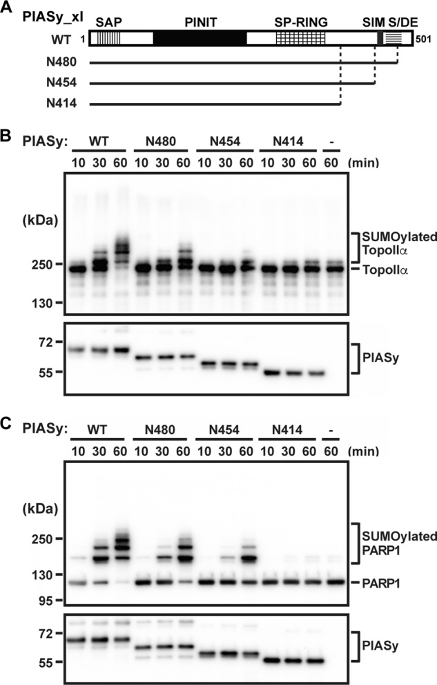
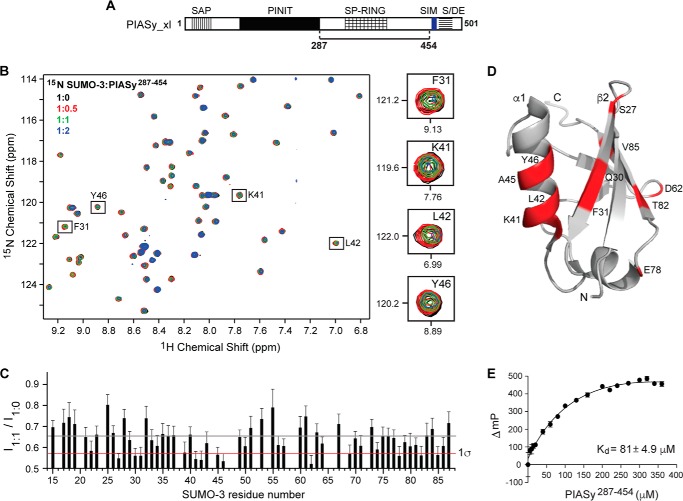
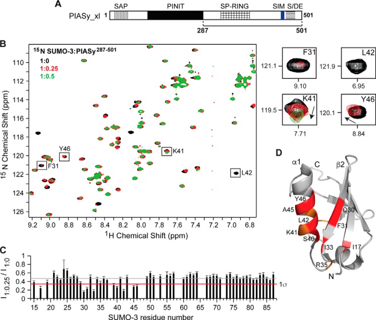
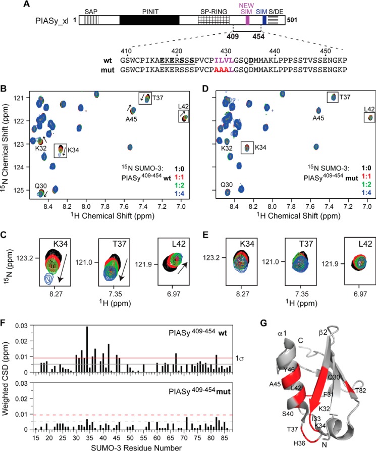
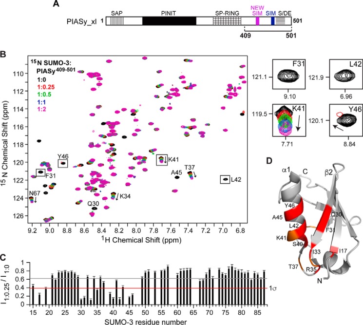
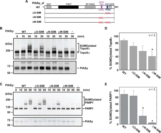
Small ubiquitin-like modifier (SUMO) conjugation is a reversible post-translational modification process implicated in the regulation of gene transcription, DNA repair, and cell cycle. SUMOylation depends on the sequential activities of E1 activating, E2 conjugating, and E3 ligating enzymes. SUMO E3 ligases enhance transfer of SUMO from the charged E2 enzyme to the substrate. We have previously identified PIASy, a member of the Siz/protein inhibitor of activated STAT (PIAS) RING family of SUMO E3 ligases, as essential for mitotic chromosomal SUMOylation in frog egg extracts and demonstrated that it can mediate effective SUMOylation. To address how PIASy catalyzes SUMOylation, we examined various truncations of PIASy for their ability to mediate SUMOylation. Using NMR chemical shift mapping and mutagenesis, we identified a new SUMO-interacting motif (SIM) in PIASy. The new SIM and the currently known SIM are both located at the C terminus of PIASy, and both are required for the full ligase activity of PIASy. Our results provide novel insights into the mechanism of PIASy-mediated SUMOylation. PIASy adds to the growing list of SUMO E3 ligases containing multiple SIMs that play important roles in the E3 ligase activity.
Keywords: PARP1; PIASy; SIM; SUMO; SUMO-interacting motif (SIM); TopoIIa; UBC9; nuclear magnetic resonance (NMR); small ubiquitin-like modifier (SUMO); sumoylation.
© 2017 by The American Society for Biochemistry and Molecular Biology, Inc.
Conflict of interest statement
The authors declare that they have no conflicts of interest with the contents of this article
Figures
Similar articles
-
Rod/Zw10 complex is required for PIASy-dependent centromeric SUMOylation.J Biol Chem. 2010 Oct 15;285(42):32576-85. doi: 10.1074/jbc.M110.153817. Epub 2010 Aug 9. J Biol Chem. 2010. PMID: 20696768 Free PMC article.
-
PIASy mediates SUMO-2/3 conjugation of poly(ADP-ribose) polymerase 1 (PARP1) on mitotic chromosomes.J Biol Chem. 2010 May 7;285(19):14415-23. doi: 10.1074/jbc.M109.074583. Epub 2010 Mar 12. J Biol Chem. 2010. PMID: 20228053 Free PMC article.
-
Site-specific inhibition of the small ubiquitin-like modifier (SUMO)-conjugating enzyme Ubc9 selectively impairs SUMO chain formation.J Biol Chem. 2017 Sep 15;292(37):15340-15351. doi: 10.1074/jbc.M117.794255. Epub 2017 Aug 7. J Biol Chem. 2017. PMID: 28784659 Free PMC article.
-
Protein interactions in the sumoylation cascade: lessons from X-ray structures.FEBS J. 2008 Jun;275(12):3003-15. doi: 10.1111/j.1742-4658.2008.06459.x. Epub 2008 May 17. FEBS J. 2008. PMID: 18492068 Review.
-
SUMO-SIM interactions: From structure to biological functions.Semin Cell Dev Biol. 2022 Dec;132:193-202. doi: 10.1016/j.semcdb.2021.11.007. Epub 2021 Nov 25. Semin Cell Dev Biol. 2022. PMID: 34840078 Review.
Cited by
-
Mechanism and function of DNA replication-independent DNA-protein crosslink repair via the SUMO-RNF4 pathway.EMBO J. 2021 Sep 15;40(18):e107413. doi: 10.15252/embj.2020107413. Epub 2021 Aug 4. EMBO J. 2021. PMID: 34346517 Free PMC article.
-
The Role of SUMO E3 Ligases in Signaling Pathway of Cancer Cells.Int J Mol Sci. 2022 Mar 26;23(7):3639. doi: 10.3390/ijms23073639. Int J Mol Sci. 2022. PMID: 35408996 Free PMC article. Review.
-
PIAS1 protects against myocardial ischemia-reperfusion injury by stimulating PPARγ SUMOylation.BMC Cell Biol. 2018 Nov 12;19(1):24. doi: 10.1186/s12860-018-0176-x. BMC Cell Biol. 2018. PMID: 30419807 Free PMC article.
-
Interaction networks of SIM-binding groove mutants reveal alternate modes of SUMO binding and profound impact on SUMO conjugation.Sci Adv. 2025 May 16;11(20):eadp2643. doi: 10.1126/sciadv.adp2643. Epub 2025 May 14. Sci Adv. 2025. PMID: 40367183 Free PMC article.
-
Inhibition of coronaviral exonuclease activity by TRIM-mediated SUMOylation.bioRxiv [Preprint]. 2025 Jul 29:2025.07.28.667286. doi: 10.1101/2025.07.28.667286. bioRxiv. 2025. PMID: 40766373 Free PMC article. Preprint.
References
MeSH terms
Substances
Associated data
- Actions
Grants and funding
LinkOut - more resources
Full Text Sources
Other Literature Sources
Miscellaneous
