. 2017 Sep;74(18):3395-3412.
doi: 10.1007/s00018-017-2523-z. Epub 2017 Apr 28.

Three Cdk1 sites in the kinesin-5 Cin8 catalytic domain coordinate motor localization and activity during anaphase

Affiliations

Three Cdk1 sites in the kinesin-5 Cin8 catalytic domain coordinate motor localization and activity during anaphase

Alina Goldstein et al. Cell Mol Life Sci. 2017 Sep.

Abstract

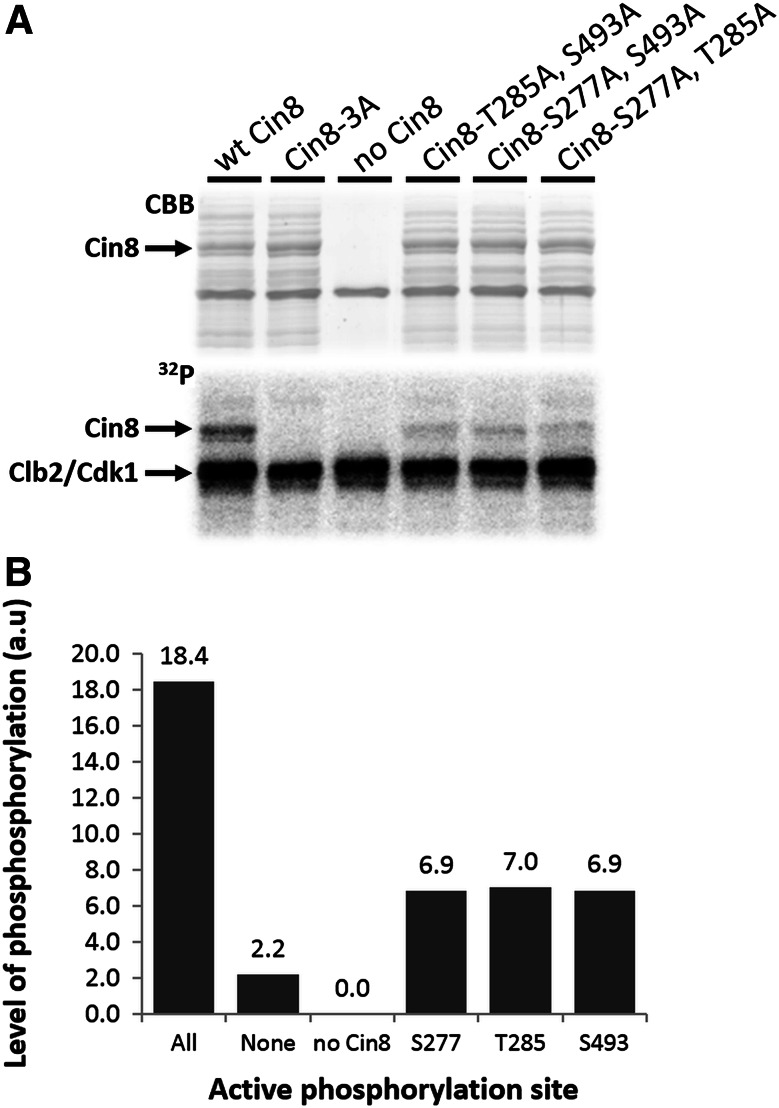
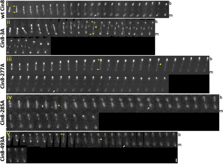
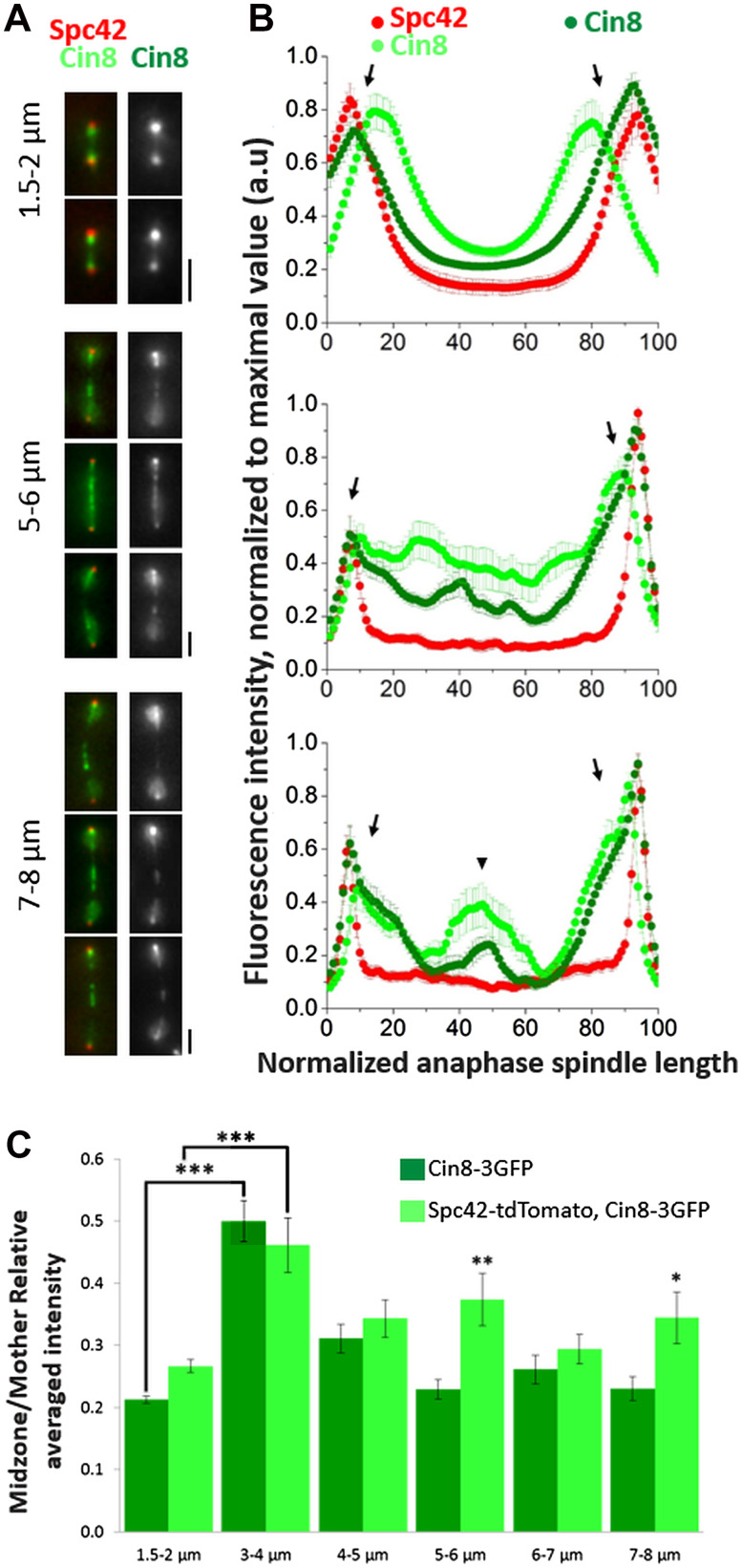
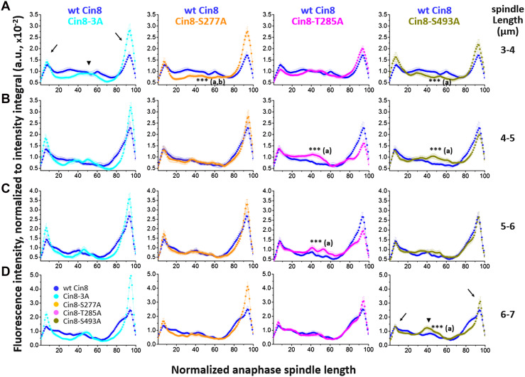
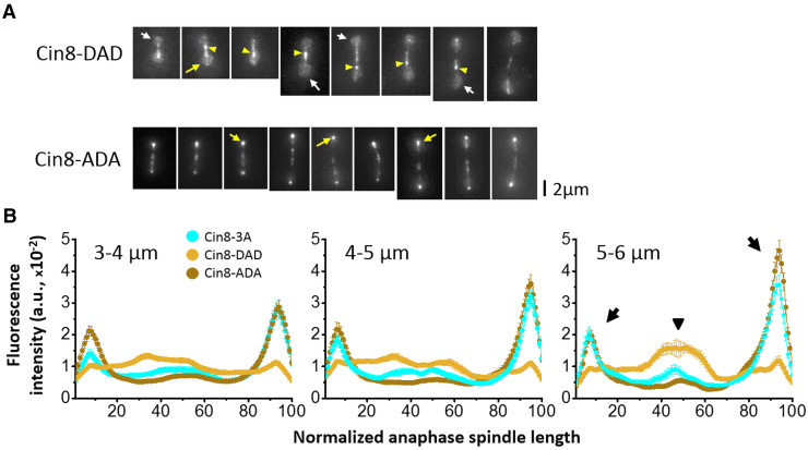
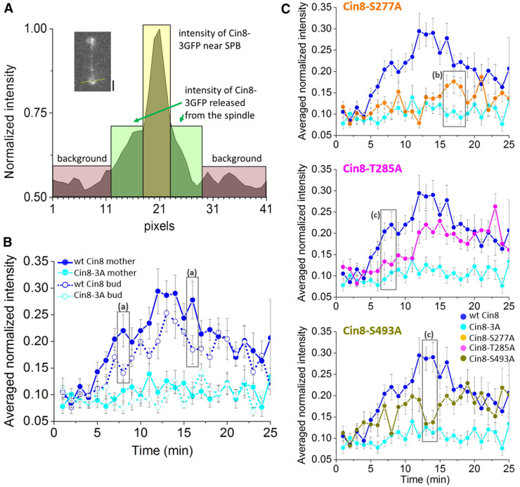
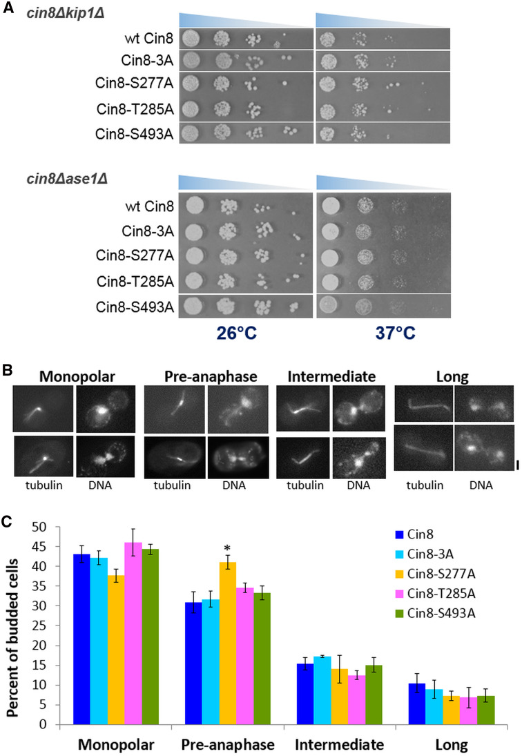
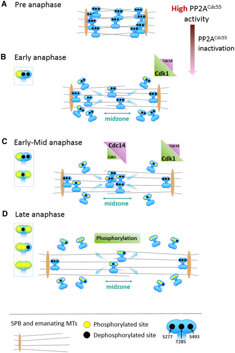
The bipolar kinesin-5 motors perform essential functions in mitotic spindle dynamics. We previously demonstrated that phosphorylation of at least one of the Cdk1 sites in the catalytic domain of the Saccharomyces cerevisiae kinesin-5 Cin8 (S277, T285, S493) regulates its localization to the anaphase spindle. The contribution of these three sites to phospho-regulation of Cin8, as well as the timing of such contributions, remains unknown. Here, we examined the function and spindle localization of phospho-deficient (serine/threonine to alanine) and phospho-mimic (serine/threonine to aspartic acid) Cin8 mutants. In vitro, the three Cdk1 sites undergo phosphorylation by Clb2-Cdk1. In cells, phosphorylation of Cin8 affects two aspects of its localization to the anaphase spindle, translocation from the spindle-pole bodies (SPBs) region to spindle microtubules (MTs) and the midzone, and detachment from the mitotic spindle. We found that phosphorylation of S277 is essential for the translocation of Cin8 from SPBs to spindle MTs and the subsequent detachment from the spindle. Phosphorylation of T285 mainly affects the detachment of Cin8 from spindle MTs during anaphase, while phosphorylation at S493 affects both the translocation of Cin8 from SPBs to the spindle and detachment from the spindle. Only S493 phosphorylation affected the anaphase spindle elongation rate. We conclude that each phosphorylation site plays a unique role in regulating Cin8 functions and postulate a model in which the timing and extent of phosphorylation of the three sites orchestrates the anaphase function of Cin8.

Keywords: Anaphase B; Cin8; Kinesin-5; Microtubules; Mitosis; Phospho-regulation.


Figures

Fig. 1
Fig. 1
In vitro Clb2-Cdk1 kinase assay of the Cin8 motor domain. a CBB staining and 32P autoradiograms of SDS-PAGE fractionation of phosphorylation reaction mixtures with five phospho-variants of Cin8: wt; 3A; S277A, T285A; S277A, S493A; and T285A, S493A. The phosphorylation reaction mixture without Cin8 served as negative control. b Qualitative level of phosphorylation. The columns quantify phosphorylation band intensity, as measured on the autoradiogram. The columns are named after the active phosphorylation sites in each mutant; “None” refers to Cin8-3A, “All” to wt Cin8
Fig. 2
Fig. 2
Localization of Cin8 phospho-variants to anaphase spindles. Time-lapse 2D-projected images of anaphase spindle in Cin8-3GFP (wt and phospho-mutants)-expressing cell. Time intervals between frames—60 s. Cin8 and its mutants, indicated on the left, were integrated into a LEU locus in cin8∆ cells (Table S1). All cells are presented with the mother cell at the bottom. Arrowheads indicate the midzone of mitotic spindle; yellow arrows indicate Cin8 concentrated at the SPB, white arrows indicate diffuse Cin8 at the nucleus; m mother cell, b bud cell. Scale bar 2 µm
Fig. 3
Fig. 3
Quantitative analysis of spindle Cin8 distribution on anaphase spindles during spindle elongation. a Representative images of spindles in cells expressing Cin8-3GFP alone or Cin8-3GFP with Spc42-tdTomato, at the different lengths indicated on the left. Tagged proteins are indicated on the top. Scale bar 2 µm. b Line-scan analysis along the spindle of cells expressing Cin8-3GFP with Spc42-tdTomato (light green and red, respectively) or Cin8-3GFP alone (dark green) at different spindle lengths, as per the representative images on the left. The fluorescence intensity along the spindle (x-axis) is measured from the mother to the bud cell and interpolated to 100 points of equal length. Spindle borders were determined either by the Spc42 fluorescence signal in cell expressing Spc42-tdTomato or by the bright Cin8 signal near the SPB, in cells expressing Cin8 alone. Arrows point to Cin8-3GFP intensity peaks at about 10 and 90% of the spindle length, while the arrowhead points to the Cin8-3GFP intensity peak at about 50% of spindle length. c Relative averaged intensity of Cin8-3GFP at the midzone (see “Materials and methods”) in cells expressing Cin8-3GFP in the different spindle lengths categories with Spc42-tdTomato (light green) or Cin8-3GFP alone (dark green). Averages ± SEM of 10–20 spindles are shown at different spindle lengths as indicated at the bottom. *P < 0.05, **P < 0.005 and ***P < 0.0005
Fig. 4
Fig. 4
Average fluorescence intensity along the spindle length of the three phospho-mutants, as compared to the fluorescence of wt Cin8. Spindle lengths categories are indicated on the right. Cin8-3GFP fluorescence intensity parallel to the spindle was measured starting at the mother cell, and divided to 100 points of equal length. The fluorescence of the background was subtracted from the Cin8-3GFP fluorescent signal, followed by normalization to the total integral of the Cin8-3GFP fluorescence signal. The signal for each mutant is compared to that of wt Cin8, indicated on top. Black arrows indicate intensity peaks at the SPBs while arrowheads point to intensity peaks at the midzone. The spindle length ranges are: a 3.0–3.9 µm; b 4.0–4.9 µm; c 5.0–5.9 µm; d 6.0–6.9 µm. For each length-segment, the average fluorescence intensity (±SEM) was determined for 14–20 cells. (a) Significance compared to wt Cin8. (b) Significance compared to Cin8-3A. ***P < 0.0005
Fig. 5
Fig. 5
Localization of Cin8 phospho-deficient/mimic variants to anaphase spindles. a Representative 2D projection of 3D stacks of fluorescent images of cells expressing 3GFP-tagged Cin8. Cin8-DAD (top) and Cin8-ADA (bottom). Arrowheads indicate Cin8 concentrating at the midzone of mitotic spindle; yellow arrows indicate Cin8 concentrated at SPB, while white arrows indicate diffuse Cin8 at the SPB. Scale bar 2 µm. b Cin8-3GFP fluorescence intensity parallel to the spindle, as in Fig. 4. The x-axis represents spindle length normalized to 100 length-segments and the y-axis represents Cin8-3GFP fluorescence intensity after the background was subtracted, followed by normalization to the total integral of the Cin8-3GFP fluorescence signal. For each length-segment, average fluorescence intensity (±SEM) was determined for 18–20 cells
Fig. 6
Fig. 6
Quantitative analysis of the detachment of Cin8 phospho-variants from mitotic spindles during anaphase. a Presentation of the method: line-scan measurements perpendicular to the spindle were performed on a single time point near the mother SPB. The corresponding image is shown at the top left; the scale bar represents 2 µm. The y-axis represents the intensity measured along the yellow line; the x-axis represents the pixels along this line. The intensity peak at 20 pixels corresponds to the high intensity of Cin8-3GFP near the SPB (yellow rectangle). Background intensity is indicated by burgundy was assigned as the background. The intensity indicated in green represents the intensity of Cin8-3GFP detached at the nucleus; this intensity was averaged and normalized per time point for each cell (see “Materials and methods”). b The average intensity of Cin8 released from the spindle of wt Cin8 (blue) and Cin8-3A (cyan) in mother (solid line) and bud (dashed line) cells versus time (min) following the onset of spindle elongation. c Anaphase release of the three phospho-mutants from the spindle, as compared to wt Cin8 and Cin8-3A. b, c At each time point, the average intensity ± SEM of 9–12 cells is presented. Gray frames contain data points with P < 0.05, (a) significance compared between wt Cin8 fluorescence in the mother and bud cells; (b) significance compared to Cin8-3A or (c) wt Cin8
Fig. 7
Fig. 7
Viability and spindle morphology in cells expressing Cin8 phospho-variants. a Growth of a shuffle cin8Δkip1Δ (top) and cin8Δase1Δ (bottom) strains on plates containing 7.5 µg/mL cyclohexamide, following transformation with CEN plasmids expressing the different Cin8 phospho-variants (indicated on the left). Cells were plated in serial dilution at 26 and 37 °C. b Representative images of fixed immunostained cells showing tubulin and DNA morphologies in the different spindle length categories: monopolar, pre-anaphase (<2 µm), intermediate anaphase (2–5 µm), and long anaphase (>5 µm) spindles. The scale bar represents 2 µm. c Distribution of spindle lengths in budded cell expressing phosphorylation mutants of Cin8. In each genotype, a total of 300 cells was counted. Budded cells were classified into four morphology classes according to spindle morphology and length, as in b. Columns and bars represent average ± SEM of three independent experiments. * P < 0.05
Fig. 8
Fig. 8
Model of Cin8 phospho-regulation during mitosis. Cin8, SPBs and MTs are illustrated. Yellow and black circles indicate phosphorylated and dephosphorylated sites (S277, T285, S493), respectively. Major phosphorylated species of Cin8 are indicated on the left of each panel. a In pre-anaphase spindles, Cin8 is dephosphorylated by PP2ACdc55 [83], resulting in its accumulation at near-SPBs regions [69]. b In early anaphase, PP2ACdc55 is inactivated by separase [83] and the local balance of Cdk1/Cdc14 near the SPBs favors Cdk1. Cin8 undergoes partial phosphorylation at the S277 site, causing Cin8 release from near-SPB sites and its relocation to spindle MTs. c During early mid anaphase, S277 and S493 undergo phosphorylation, causing additional relocation of Cin8 from the SPBs to the midzone. Release of the Cdc14 phosphatase by the FEAR pathway [87, 89] likely shifts the local Cdk1/Cdc14 balance at the midzone towards Cdc14, keeping Cin8 (and Ase1, not shown) mainly de-phosphorylated at the T285 and S493 sites. This promotes Cin8 attachment to midzone MTs allowing spindle elongation during the first, fast phase of anaphase B. d In late anaphase, Cin8 is detached from the midzone and SPBs as a result of phosphorylation at all three sites in the motor domain [56]

Similar articles

Cited by

References

    1. Maddox P, Straight A, Coughlin P, Mitchison TJ, Salmon ED. Direct observation of microtubule dynamics at kinetochores in Xenopus extract spindles: implications for spindle mechanics. J Cell Biol. 2003;162(3):377–382. doi: 10.1083/jcb.200301088. - DOI - PMC - PubMed
    1. Hoyt MA, He L, Loo KK, Saunders WS. Two Saccharomyces cerevisiae kinesin-related gene products required for mitotic spindle assembly. J Cell Biol. 1992;118(1):109–120. doi: 10.1083/jcb.118.1.109. - DOI - PMC - PubMed
    1. Hagan I, Yanagida M. Kinesin-related cut7 protein associates with mitotic and meiotic spindles in fission yeast. Nature. 1992;356(6364):74–76. doi: 10.1038/356074a0. - DOI - PubMed
    1. Heck MM, Pereira A, Pesavento P, Yannoni Y, Spradling AC, Goldstein LS. The kinesin-like protein KLP61F is essential for mitosis in Drosophila. J Cell Biol. 1993;123(3):665–679. doi: 10.1083/jcb.123.3.665. - DOI - PMC - PubMed
    1. Blangy A, Lane HA, d’Herin P, Harper M, Kress M, Nigg EA. Phosphorylation by p34cdc2 regulates spindle association of human Eg5, a kinesin-related motor essential for bipolar spindle formation in vivo. Cell. 1995;83(7):1159–1169. doi: 10.1016/0092-8674(95)90142-6. - DOI - PubMed

MeSH terms