. 2017 Jun;22(2):141-157.
doi: 10.1007/s10911-017-9379-6. Epub 2017 Apr 28.

Developmental Expression of Claudins in the Mammary Gland

Affiliations

Developmental Expression of Claudins in the Mammary Gland

Heidi K Baumgartner et al. J Mammary Gland Biol Neoplasia. 2017 Jun.

Abstract

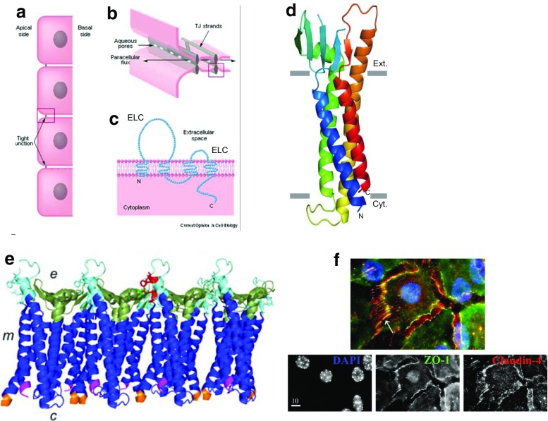
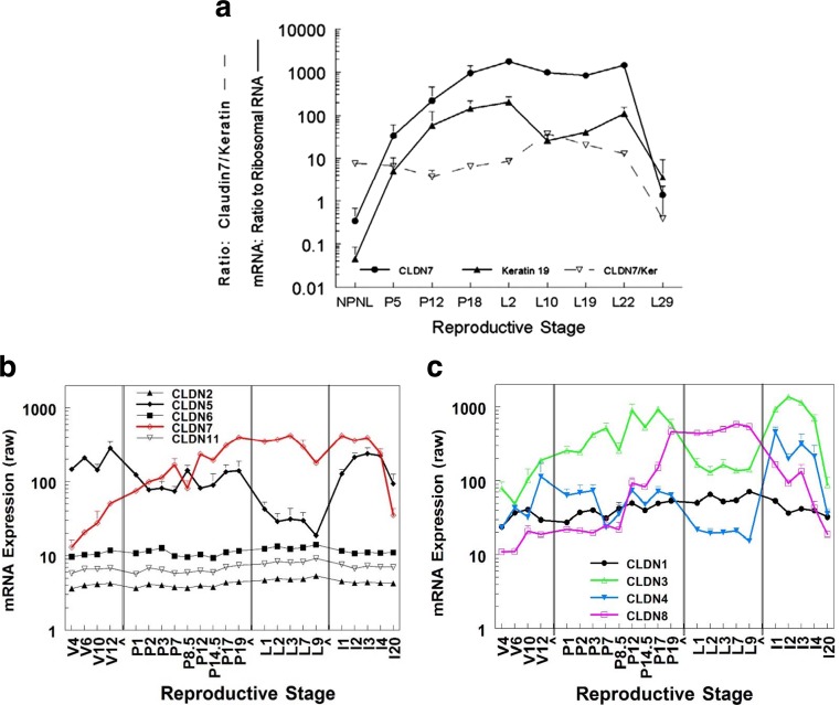
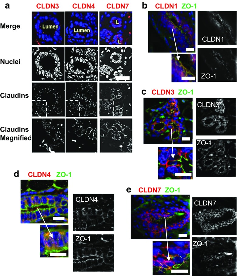
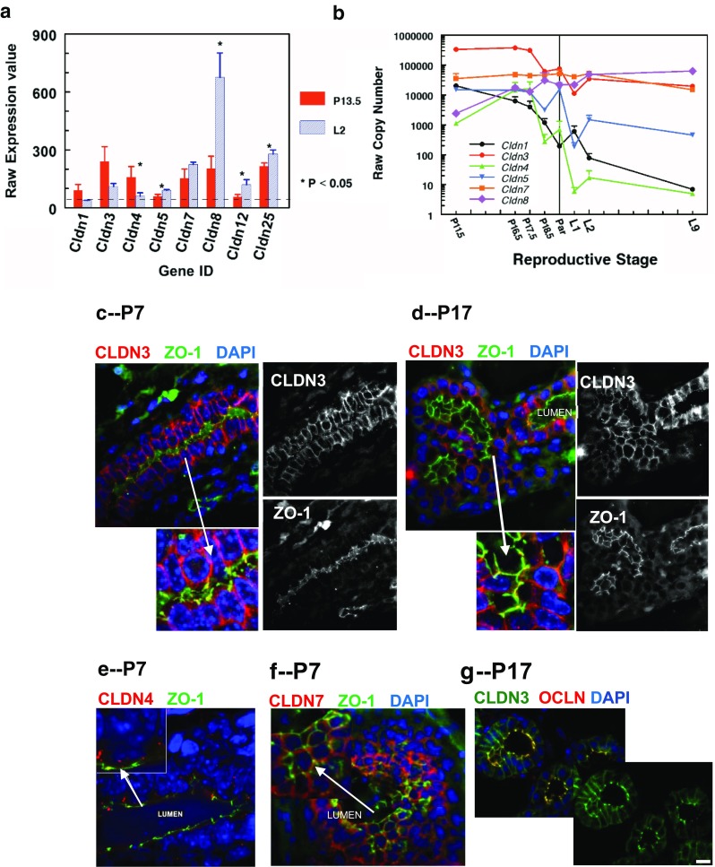
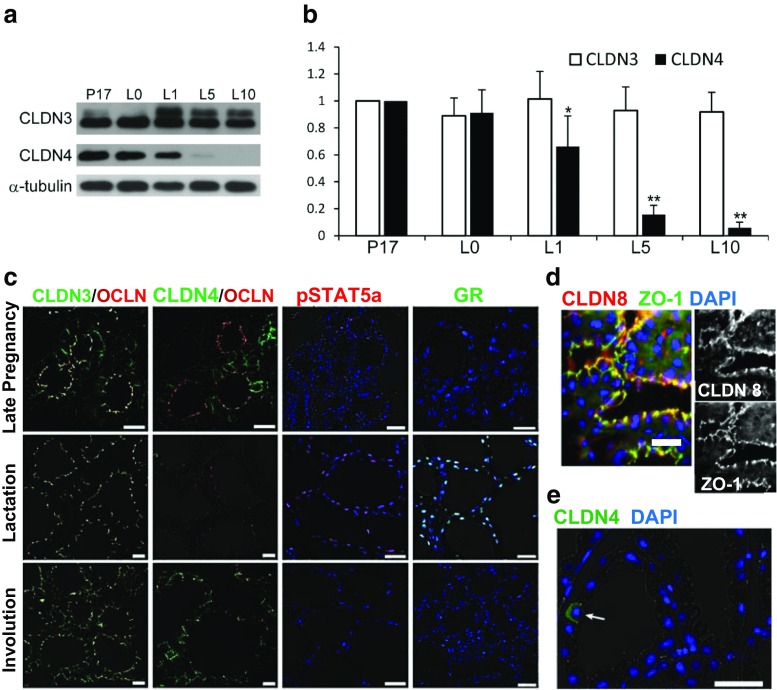
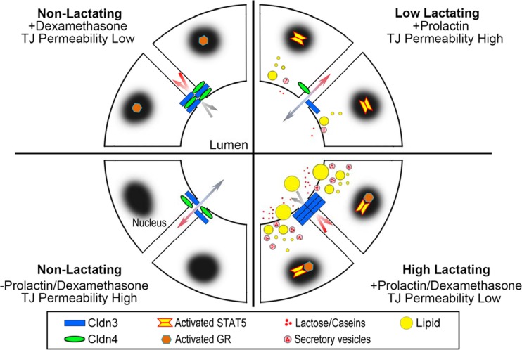
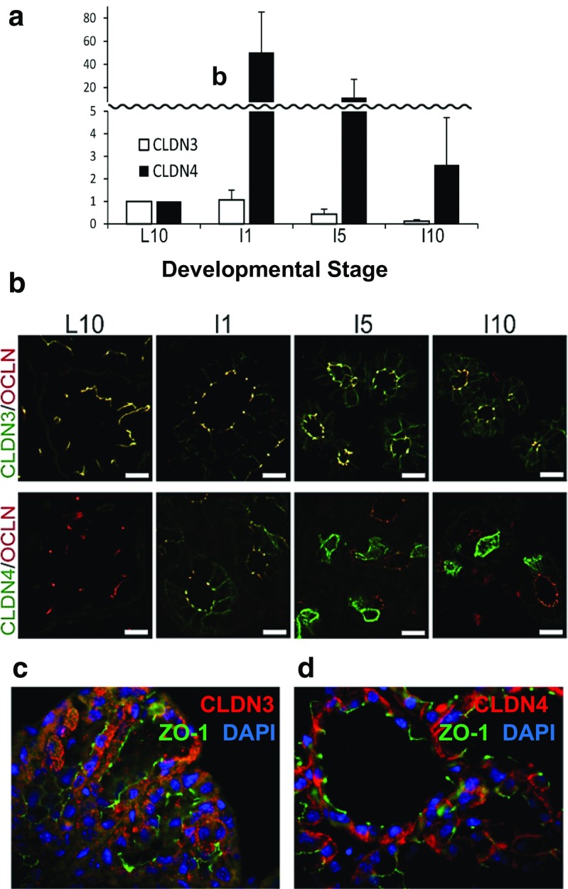
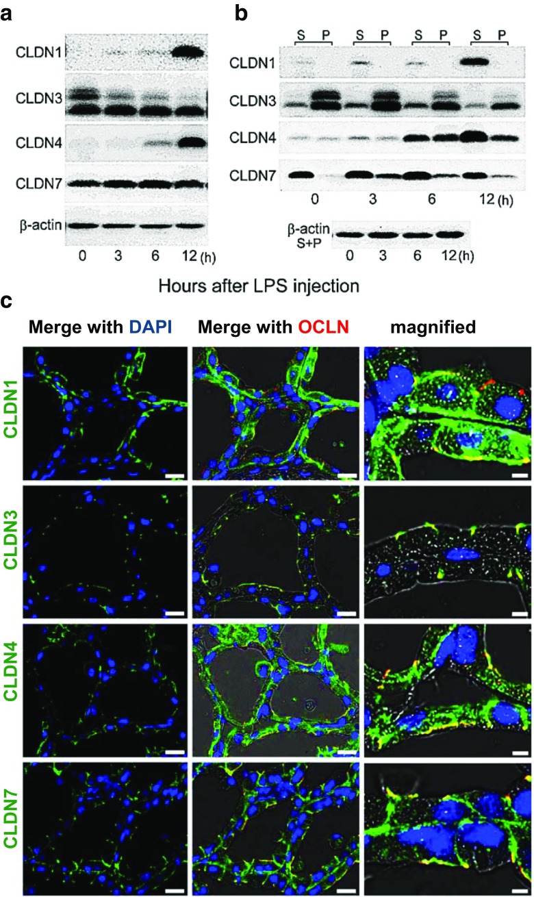
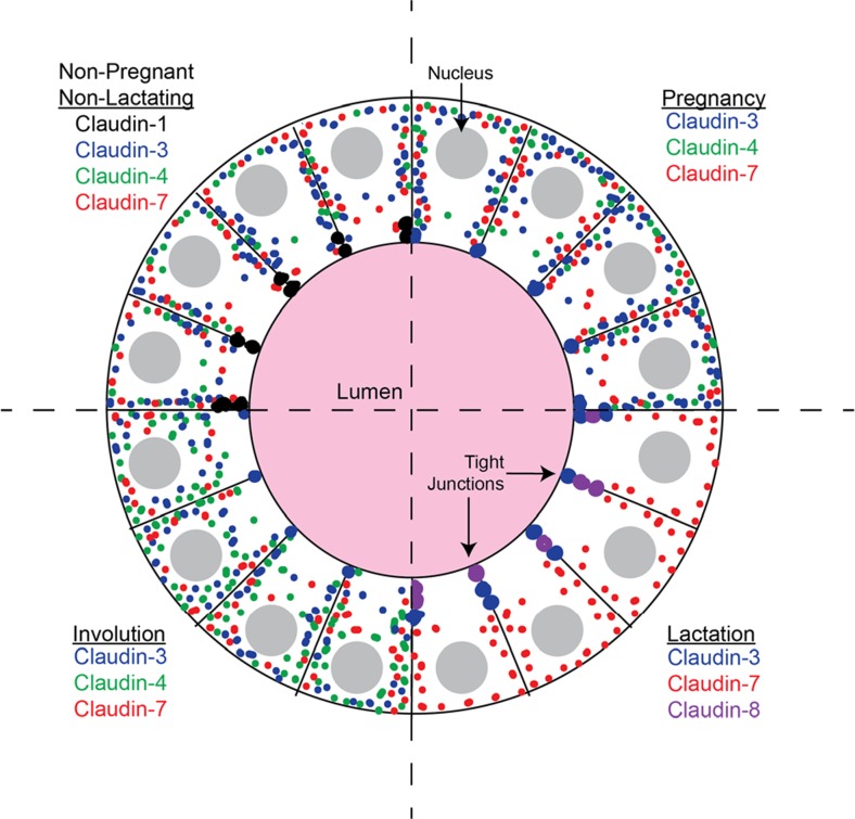
Claudins are a large family of membrane proteins whose classic function is to regulate the permeability of tight junctions in epithelia. They are tetraspanins, with four alpha-helices crossing the membrane, two extracellular loops, a short cytoplasmic N-terminus and a longer and more variable C-terminus. The extracellular ends of the helices are known to undergo side-to-side (cis) interactions that allow the formation of claudin polymers in the plane of the membrane. The extracellular loops also engage in head-to-head (trans) interactions thought to mediate the formation of tight junctions. However, claudins are also present in intracellular structures, thought to be vesicles, with less well-characterized functions. Here, we briefly review our current understanding of claudin structure and function followed by an examination of changes in claudin mRNA and protein expression and localization through mammary gland development. Claudins-1, 3, 4, 7, and 8 are the five most prominent members of the claudin family in the mouse mammary gland, with varied abundance and intracellular localization during the different stages of post-pubertal development. Claudin-1 is clearly localized to tight junctions in mammary ducts in non-pregnant non-lactating animals. Cytoplasmic puncta that stain for claudin-7 are present throughout development. During pregnancy claudin-3 is localized both to the tight junction and basolaterally while claudin-4 is found only in sparse puncta. In the lactating mouse both claudin-3 and claudin-8 are localized at the tight junction where they may be important in forming the paracellular barrier. At involution and under challenge by lipopolysaccharide claudins -1, -3, and -4 are significantly upregulated. Claudin-3 is still colocalized with tight junction molecules but is also distributed through the cytoplasm as is claudin-4. These largely descriptive data provide the essential framework for future mechanistic studies of the function and regulation of mammary epithelial cell claudins.

Keywords: Claudin-1; Claudin-3; Claudin-4; Claudin-7; Claudin-8; Extra-junctional claudin; Infection; Involution; Mammary gland.


Conflict of interest statement

All authors confirm no non-financial conflicts of interest.

Figures

Fig. 1
Fig. 1
Localization and molecular structure of claudins. a Model mucosal epithelial layer showing apical location of tight junctions. b Aqueous pores formed by some claudins in the tight junction provide a pathway for paracellular ionic flux. c. Diagrammatic view of a claudin molecule in the plasma membrane showing the four transepithelial regions, the two extracellular loops (ECS) and the cytoplasmic N and C termini. a,b,c, from [23], Current Opinion in Cell Biology. d Detailed crystal structure of the claudin-15 molecule showing its orientation in the plasma membrane, represented by grey rectangles; image from ref. [76], Science; used by permission. Transmembrane segments (TM) are shown as alpha-helices with TM1 in blue, TM2 in green with the four beta-sheets of extracellular loop 1 between them. TM3 is yellow with the helix extending beyond the membrane into the interstitial space; TM4 is red and ECL2 is between TM3 and TM4 and contains one beta-sheet. The C-terminus of the molecule was removed to facilitate crystallization and is not shown. e Claudins are thought to polymerize in the membrane via interactions between amino acids on membrane adjacent portions of ECL1 (bright green) and ECL2 (red). Image shows claudin-15 polymer with extracellular side (e), cytoplasmic side (c), and membrane (m) Image from [10], The Biochenical Journal. f Immunofluorescent localization of claudin-4 in subconfluent cultured primary mammary epithelial cells. Upper image, colocalization of claudin-4 (red) with the tight junction protein ZO-1 (green) and nuclei (blue). Claudin-4 is colocalized with ZO-1 in the tight junctions; it is also present as cytoplasmic puncta and in cellular projections that show no sign of ZO-1 staining. Image from ref. [18], BMC Cell Biology
Fig. 2
Fig. 2
Developmental expression of claudin mRNA in the murine mammary gland. a Developmental expression of claudin-7 compared to expression of the epithelial keratin 19 in glands of CD1 mice using real time PCR. Graph from Reference [29] Breast Cancer Research. b, c. Time course data obtained from Affymetrix arrays (MG-U74Av2--able to hybridize 12,000 genes) performed in the Gusterson [48] and Neville [49] laboratories. Data are combined to demonstrate how these claudins vary through the reproductive cycle. V, glands isolated from non-pregnant non-lactating mice at 4, 6, 10 and 12 weeks after birth. P (pregnancy), glands isolated 0, 1, 2, 3, 7, 10.5, 12, 14.5, 17 and 19 days after mating. Birth occurs about 19.5 days after mating in these two strains. L (lactating), glands isolated at 1, 2, 3, 7 and 9 days after parturition. I, (involution) glands 1, 2, 3, 4 and 20 days after pup removal on day 8 ([48]) or 10 ([49]) of lactation. b. Expression of claudins-2, 5, 6, and 11 compared to that of claudin-7. c.Expression of claudins-1, −3, −4, and −8 as indicated
Fig. 3
Fig. 3
Immunofluorescence analysis of claudins in the mammary gland of the non-pregnant, non-lactating mouse. a. Immunofluorescence analysis of Claudins-3, −4, and −7 in mammary glands of the FVB mouse highlighting the punctate nature of the stain localized in the ductal cells. b, c, d. Immunofluorescent images of ductal sections mammary glands from CD1 virgin mice stained for claudins-1, −3, −4 respectively (red stain) and the tight junction protein ZO-1 (green). b. Claudin-1 overlapped heavily with ZO-1, appearing yellow in merged images, indicating association with tight junctions in this ductal section. c. Claudin-3 was heavily stained along lateral and basal borders but showed little overlap with ZO-1. d. Claudin-4 was observed as sparse cytoplasmic puncta, again with little overlap with ZO-1. e. Claudin-7 was localized along the basal and lateral border of the ductal cells in the CD1 mouse with little or no overlap with ZO-1 See also ref. [29]. Scale bars, 20 μm. See Methods section for tissue preparation and staining methods for all these figures
Fig. 4
Fig. 4
Expression and localization of claudins in pregnancy. a. Microarray analysis of claudin mRNA in MECs isolated from pregnant (day 13.5) and lactating (day 2) FVB mice [58]. MECs were isolated from the 4th mammary gland according to the techniques of Rudolph and colleagues [58]. mRNA expression was analyzed using Affymetrix MoGene_1_0-st-v1 chip arrays [77]. Raw expression values for all claudins with expression values above 40 (dotted line) at pregnancy day 13.5 (red) or lactation day 2 (blue) are shown. * Expression significantly different between pregnancy and lactation, P < 0.05 b. mRNA expression of claudins 1, 3, 4, 5, 7 and 8 during the transition from pregnancy to lactation by quantitative real time PCR using lysates of whole mammary glands (See Methods, Fig. 4). d,e. Immunofluorescence localization of claudins-3, −4 and −7 in mammary glands from CD1 pregnant mice (Neville laboratory, see Methods section). c,e,f. Pregnancy day 7. Similar results were observed in both formalin-fixed (shown) and frozen (not shown) sections. d,g. Pregnancy day 17. g. Localization of claudin-3 in the mammary gland from the ICR mouse; in this strain claudin-3 was localized both basolaterally and with occludin at pregnancy day 17 [43]. c-g. Scale bars, 20 μm
Fig. 5
Fig. 5
Protein expression and localization for claudins in the mammary gland of mice in mid-lactation. a Western blots for claudins-3 and -4 in the lactating gland at days 0, 1, 5, and 10 compared to late pregnancy (P17). Alpha-tubulin was the loading control. b Quantitation of Western blots for claudin-3 and claudin-4 compared to late pregnancy (P17). Significantly different from P17, *P < 0.05; **P < 0.01. c. Subcellular localization of claudins-3 and -4, pSTAT5a and the glucocorticoid receptor (GR) in the murine mammary gland at day 10 of lactation. Paraffin sections were immunostained with antibodies to the relevant claudin (green), occludin (red), pSTAT5a (red) and GR (green). Blue; nuclei stained with DAPI. d Immunofluorescence analysis of claudin-8 from the mammary gland of a day 2 lactating mouse. e. Rare occurrence of basolateral CLDN4 in a mammary alveolar cell of a lactating mouse. Scale bars; 20 μm. Images a and b based on ref. [43], image c based on Ref [64] and image e from Ref [31] Plos One, all ICR mice. Image d, Neville laboratory (CD1 mouse; see methods)
Fig. 6
Fig. 6
Model for hormonal regulation of claudin-3 and claudin-4 in the lactating mammary gland. Data from cultures of mouse mammary epithelial cells show that claudin-3 and claudin-4 respond to prolactin and glucocorticoids, major regulators of secretory activation in a manner similar to casein and lipid secretion [43]. See text for details
Fig. 7
Fig. 7
Claudins in mammary involution. a Levels of claudins-3 and -4 protein from Western blots during involution. Pups were removed from day 10 lactating ICR mice. Dams were sacrificed at day 10 of lactation and 1, 5 and 10 days later. Proteins from minced mammary glands were electrophoresed and visualized with appropriate antibodies (ThermoFisher) as described [43]. b Immunofluorescence of claudins-3 and -4 in the samples from panel a compared to localization of the tight junction protein, occludin (OCLN). Images from Kobayashi lab after ref. [43]. c,d Higher power images from sections of mammary glands from 10 day lactating FVB mice sacrificed 2 days after pup removal and stained using antibodies for claudin-3, claudin-4 (claudins in red) and the tight junction protein ZO-1 (green). Nuclei stained with DAPI (blue) Significant cytoplasmic stain can be observed for both claudins with little or no overlap with ZO-1. See methods for preparation of these images from the Neville laboratory
Fig. 8
Fig. 8
Effect of LPS on amount, type and localization of claudin protein. a. Western blot of claudins-1, −3, −4, and −7 in 10 day lactating gland 0, 3, 6, and 12 h after injection of E. coli lipopolysaccharide (LPS) into the teat canal of the fourth mammary gland of ICR mice. b. Western blot of claudins in Triton-X soluble (S) and insoluble (P) fractions of mammary gland lysates after LPS injection. c. Immunofluorescence analysis of claudins in the mammary gland 12 h after injection of LPS. Figures from ref. [31] Plos One
Fig. 9
Fig. 9
Model for distribution of claudins between cytoplasm and tight junctions at different stages of mammary development. See text for explanation

References

    1. Furuse M, Fujita K, Hiiragi T, Fujimoto K, Tsukita S. Claudin-1 and -2: novel integral membrane proteins localizing at tight junctions with no sequence similarity to occludin. J Cell Biol. 1998;141(7):1539–1550. doi: 10.1083/jcb.141.7.1539. - DOI - PMC - PubMed
    1. Kolosov D, Bui P, Chasiotis H, Kelly SP. Claudins in teleost fishes. Tissue barriers. 2013;1(3) doi: 10.4161/tisb.25391. - DOI - PMC - PubMed
    1. Mineta K, Yamamoto Y, Yamazaki Y, Tanaka H, Tada Y, Saito K, et al. Predicted expansion of the claudin multigene family. FEBS Lett. 2011;585(4):606–612. doi: 10.1016/j.febslet.2011.01.028. - DOI - PubMed
    1. Furuse M, Sasaki H, Fujimoto K, Tsukita S. A single gene product, claudin-1 or −2, reconstitutes tight junction strands and recruits occludin in fibroblasts. J Cell Biol. 1998;143(2):391–401. doi: 10.1083/jcb.143.2.391. - DOI - PMC - PubMed
    1. Furuse M, Hata M, Furuse K, Yoshida Y, Haratake A, Sugitani Y, et al. Claudin-based tight junctions are crucial for the mammalian epidermal barrier: a lesson from claudin-1-deficient mice. J Cell Biol. 2002;156(6):1099–1111. doi: 10.1083/jcb.200110122. - DOI - PMC - PubMed

Publication types