Review
. 2017 May 1;14(1):12.
doi: 10.1186/s12987-017-0061-6.

NIH workshop report on the trans-agency blood-brain interface workshop 2016: exploring key challenges and opportunities associated with the blood, brain and their interface

Affiliations
Review

NIH workshop report on the trans-agency blood-brain interface workshop 2016: exploring key challenges and opportunities associated with the blood, brain and their interface

Margaret J Ochocinska et al. Fluids Barriers CNS. .

Abstract

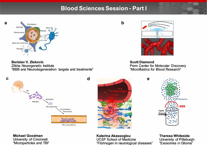
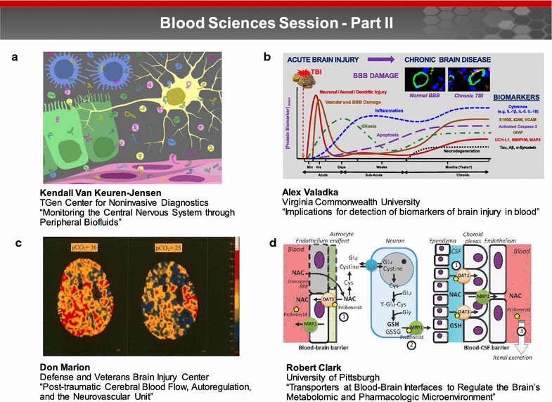
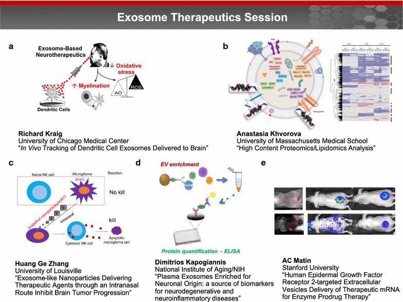
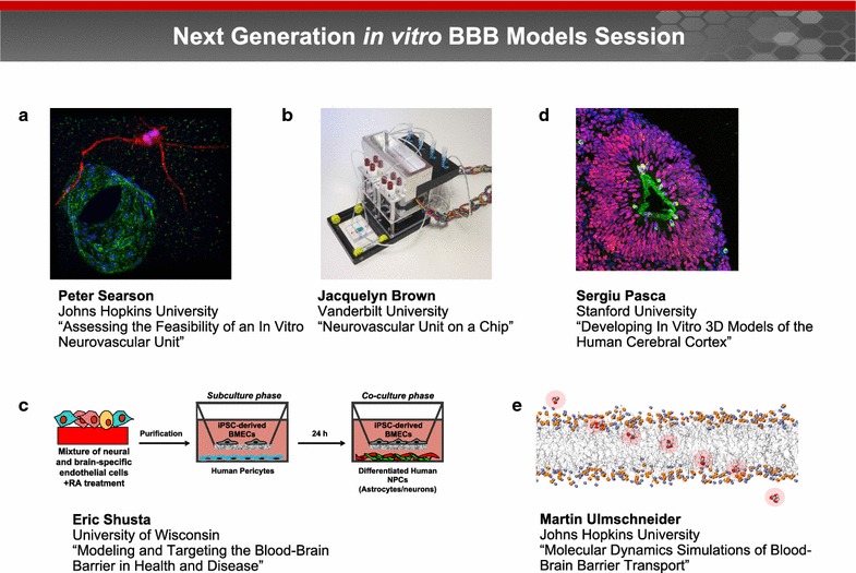
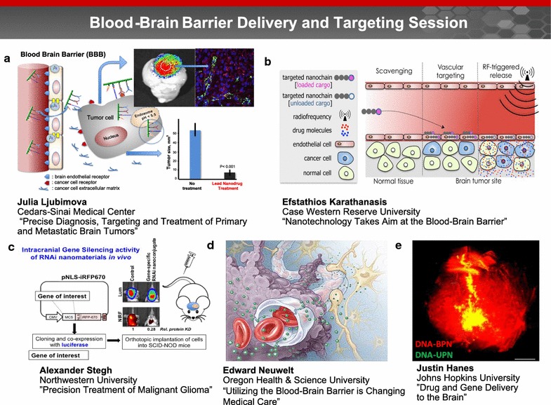
A trans-agency workshop on the blood-brain interface (BBI), sponsored by the National Heart, Lung and Blood Institute, the National Cancer Institute and the Combat Casualty Care Research Program at the Department of Defense, was conducted in Bethesda MD on June 7-8, 2016. The workshop was structured into four sessions: (1) blood sciences; (2) exosome therapeutics; (3) next generation in vitro blood-brain barrier (BBB) models; and (4) BBB delivery and targeting. The first day of the workshop focused on the physiology of the blood and neuro-vascular unit, blood or biofluid-based molecular markers, extracellular vesicles associated with brain injury, and how these entities can be employed to better evaluate injury states and/or deliver therapeutics. The second day of the workshop focused on technical advances in in vitro models, BBB manipulations and nanoparticle-based drug carrier designs, with the goal of improving drug delivery to the central nervous system. The presentations and discussions underscored the role of the BBI in brain injury, as well as the role of the BBB as both a limiting factor and a potential conduit for drug delivery to the brain. At the conclusion of the meeting, the participants discussed challenges and opportunities confronting BBI translational researchers. In particular, the participants recommended using BBI translational research to stimulate advances in diagnostics, as well as targeted delivery approaches for detection and therapy of both brain injury and disease.

Keywords: Blood–brain barrier; Cancer; Delivery; Exosomes; Extracellular vesicles; Neurodegeneration; Therapeutics; Traumatic brain injury.


Figures

Fig. 1
Fig. 1
Blood sciences session—Part I. Part I of the blood sciences session focused on the physiology of the blood and neurovascular unit, biofluid-based molecular markers, and extracellular vesicles associated with brain injury and disease progression. The presentations explored how these entities are transported between brain and blood and can be employed to evaluate injury states or deliver therapeutics. a Endothelial cells, pericytes, neurons and glia constitute an interconnected functional cellular network, collectively referred to as the neurovascular unit. Dr. Berislav Zlokovic discussed how defects in blood vessels and the neurovascular unit can lead to BBB breakdown and neurodegeneration (image reprinted with permission from [107]). b The Diamond laboratory uses a combination of microfluidics, patient-specific high throughput phenotyping, and systems biology to quantify blood function in the hemodynamic and pharmacological context of thrombosis and hemostasis. Image courtesy of Dr. Diamond. c Dr. Michael Goodman discussed how traumatic brain injury (TBI) induces systemic alterations in the aggregation of platelets aggregation and as well as the generation and function of microparticles (MP). Image courtesy of Dr. Goodman. d Dr. Katerina Akassoglou identified fibrinogen as a novel molecular link between blood–brain barrier disruption, activation of CNS innate immunity, and neurodegeneration. Image courtesy of Dr. Akassoglou. e Dr. Theresa Whiteside noted that genetic and molecular cargo of exosomes found in plasma of glioma patients resembles that of tumor cells and suggested that tumor derived exosomes hold promise as biomarkers of prognosis, as a source of liquid biopsy. Image courtesy of Dr. Whiteside
Fig. 2
Fig. 2
Blood sciences session—Part II. Tools and technologies are currently being developed to improve capabilities for assessment and monitoring of CNS trauma to: (1) explore the relationship among the post-traumatic cerebral blood, autoregulation and the neurovascular unit; and (2) leverage different transporters at the BBIs to regulate the brain metabolomics and pharmacologic microenvironment. a Dr. Kendall Van Keuren-Jensen discussed the need for accessible biomarkers to allow more frequent monitoring of the CNS and the potential to improve patient care. The presence of RNA in body fluids and its relative stability when transported via EVs or carrier proteins has captured significant attention as a source for biomarker discovery. Image courtesy of the NIH exRNA Communication Program. b Dr. Alex Valadka noted that differences in markers released by neuronal and glial cells may make possible the diagnosis of specific subtypes of TBI based on distinct patterns and ratios of glial and neuronal markers. Image courtesy of Dr. Valadka. c Brain trauma can result in an immediate decrease in the cerebral blood flow, resulting in loss, or fluctuations in multiple physiological control systems including blood pressure and chemical autoregulation. Image courtesy of Dr. Marion. d Dr. Robert Clark reported on the importance of membrane transporters at the BBI. These membrane transporters are responsible for efflux of exogenous substrates (e.g. drugs) at the BBB and CSF-blood barriers, impacting the brain’s microenvironment. Image courtesy of Dr. Clark
Fig. 3
Fig. 3
Exosome therapeutics session. Exosomes, the smallest (30–150 nm) type of cell-derived extracellular vesicles, are present in all body fluids and serve as carriers of information across the body in both health and disease. This session focused on the emerging role of exosomes, in particular how they can be harnessed for both diagnostic and targeted drug delivery applications. a Dr. Richard Kraig reported that interferon gamma (IFN)-stimulated dendritic cells (SDCs) release SDC-derived exosomes (SDC-exos) containing specific miRNAs which promote myelination and reduce oxidative stress when administered to brain slice cultures. Image courtesy of Dr. Kraig. b Dr. Anastasia Khvorova and coworkers are examining the surface lipid and protein composition of EV membranes (left image modified from [108]) through high content proteomics and lipidomics analysis to better understand how these elements contribute to EV function. Right image courtesy of Dr. Khvorova. c Dr. Huang Ge Zhang described a novel approach for grapefruit derived nanovector (GNV)-mediated intranasal delivery of RNA in general and therapeutic miR17 specifically to brain tumor cells as a proof of concept. miR17-mediated downregulation of MHC1 expressed on tumor cells leads to activation of Natural Killer (NK) cells and targeting of tumor cells. Image courtesy of Dr. Zhang. d Dr. Dimitrios Kapogiannis and coworkers developed a method for enriching peripheral blood EVs of neuronal origin using L1 cell adhesion molecule (L1CAM) immunoprecipitation. In case-controlled studies, L1CAM + EVs showed diagnostic differences for AD, and perhaps fronto-temporal dementia, multiple sclerosis, and TBI. Image courtesy of Dr. Kapogiannis. e By using EVs as the “body’s antigen delivery system” for targeting a novel prodrug 6-chloro-9-nitro-5-oxo-5H-benzo(a)phenoxazine (CNOB)/ChrR6 regimen specifically to HER2 positive cancer, the cytotoxic product of this regimen, 9-Amino-6-chloro-5H-benzo(a)phenoxazine-5-one (MCHB), can be visualized noninvasively in living mice. Image reprinted with permission from [58]
Fig. 4
Fig. 4
Next generation in vitro BBB models session. Next generation BBB models span organ-on-a-chip devices and models exploiting organogenesis, microfluidics, 3D printing, and self-organization. These models are enabled by advances in human brain-specific cell lines and tissue engineering, particularly in 3D co-culture and matrix materials. a Dr. Peter Searson noted that stem cell technology has the potential to overcome the limitations of animal-derived cells and immortalized cell lines, and provide a source of human brain-specific microvascular endothelial cells, astrocytes, and pericytes. induced pluripotent stem cells (iPSCs) can be used to generate human brain microvascular endothelial cells (hBMECs) for developing perfusable brain capillary networks that capture the important physical and biological characteristics of the BBB in a physiologically relevant geometry. Image courtesy of Dr. Searson. b Dr. Jacquelyn Brown and colleagues developed a transwell assay that consists of a monolayer of endothelial cells seeded on a porous membrane that is separated into apical and basolateral chambers on either side of the membrane. These platforms have perfusable vascular and brain chambers on either side of the membrane that can be used to measure solute permeability and asses brain penetration. Image courtesy of Dr. Brown. c Dr. Eric Shusta reported how endothelial cells generated from human pluripotent stem cells (hPSCs) can be programmed to possess many BBB attributes, including well-organized tight junctions, polarized efflux transport, and nutrient transporter expression. Image courtesy of Dr. Shusta. d Dr. Sergiu Pasca discussed how spheroids of pluripotent stem cells (PSCs) can be programmed to form structures that resemble the human cerebral cortex. Image courtesy of Dr. Pasca. e Advances in computational methods have enabled a new generation of tools for modeling BBB transport. Dr. Martin Ulmschneider described how these models can simulate spontaneous transmembrane diffusion of small molecules using unbiased long timescale atomic detail molecular dynamics (MD) techniques. Image courtesy of Dr. Ulmschneider
Fig. 5
Fig. 5
Blood brain barrier delivery and targeting session. Drug delivery through the BBB to the brain can be enhanced utilizing nanoparticles that are designed to exhibit BBB targeting and/or penetrating capabilities. Evidence of these nanoparticles across the BBB can be accessed via in vivo imaging. a Dr. Julia Ljubimova presented a novel nanotechnology which can overcome the BBB for precise diagnosis, targeting and treatment of primary and metastatic brain tumors. Use of systemically administered novel nanobiopolymers based on a combination of a polymalic acid platform (Polycefin™ family of nano agents), nano drugs and imaging agents, dramatically reduced tumor size by 90% and normalized brain cancer vasculature. Image courtesy of Dr. Ljubimova. b Dr. Karathanasis’ group has developed a new class of multicomponent chain-like nanoparticles, termed nanochains. Due to enhanced site-specific targeting and radiofrequency-triggered drug release, the nanochains facilitate effective delivery of drugs across the BBB into hard-to-reach brain tumors (image reprinted with permission from [91]). c Dr. Alexander Stegh and his team developed RNAi-based Spherical Nucleic Acids (SNAs) for the treatment of Glioblastoma multiforme (GBM). Upon IV administration, SNAs cross the BBB and disseminate within intracranial patient-derived xenografts and genetically engineered mouse model tumors. Image courtesy of Dr. Stegh. d Dr. Edward Neuwelt emphasized that advances toward penetrating the BBB must consider both normal and abnormal function, as well as the entire neurovascular unit (illustrated here). Image courtesy of Dr. Neuwelt. e Dr. Justin Hanes presented a nanoparticle-based platform for drug delivery to the brain. Brain-penetrating DNA nanoparticles (red color) spread throughout the entire rat striatum, as compared to standard DNA nanoparticles (yellow color) that do not spread as well. Image courtesy of Dr. Panagiotis Mastorakos and the work is a collaboration between Dr. Hanes’ research group and that of Prof. Jung Soo Suk

Similar articles

Cited by

References

    1. van der Pol E, Boing AN, Harrison P, Sturk A, Nieuwland R. Classification, functions, and clinical relevance of extracellular vesicles. Pharmacol Rev. 2012;64(3):676–705. doi: 10.1124/pr.112.005983. - DOI - PubMed
    1. Hynynen K, McDannold N, Vykhodtseva N, Jolesz FA. Noninvasive MR imaging-guided focal opening of the blood–brain barrier in rabbits. Radiology. 2001;220(3):640–646. doi: 10.1148/radiol.2202001804. - DOI - PubMed
    1. McDannold N, Arvanitis CD, Vykhodtseva N, Livingstone MS. Temporary disruption of the blood–brain barrier by use of ultrasound and microbubbles: safety and efficacy evaluation in rhesus macaques. Cancer Res. 2012;72(14):3652–3663. doi: 10.1158/0008-5472.CAN-12-0128. - DOI - PMC - PubMed
    1. Zhao Z, Nelson AR, Betsholtz C, Zlokovic BV. Establishment and dysfunction of the blood–brain barrier. Cell. 2015;163(5):1064–1078. doi: 10.1016/j.cell.2015.10.067. - DOI - PMC - PubMed
    1. Bell RD, Winkler EA, Singh I, Sagare AP, Deane R, Wu Z, Holtzman DM, Betsholtz C, Armulik A, Sallstrom J, et al. Apolipoprotein E controls cerebrovascular integrity via cyclophilin A. Nature. 2012;485(7399):512–516. - PMC - PubMed

MeSH terms