. 2017 Jun:107:41-51.
doi: 10.1016/j.yjmcc.2017.04.004. Epub 2017 Apr 27.

Non-nuclear estrogen receptor alpha activation in endothelium reduces cardiac ischemia-reperfusion injury in mice

Affiliations

Non-nuclear estrogen receptor alpha activation in endothelium reduces cardiac ischemia-reperfusion injury in mice

Sara Menazza et al. J Mol Cell Cardiol. 2017 Jun.

Abstract

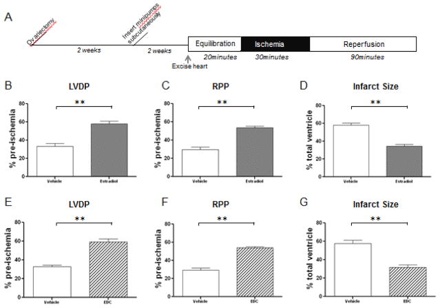
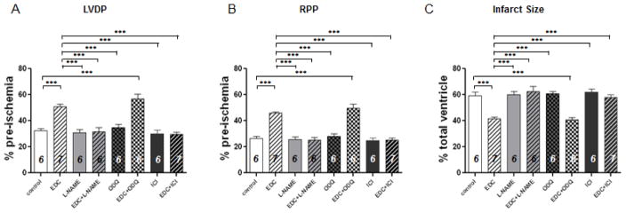
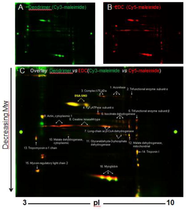
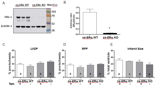
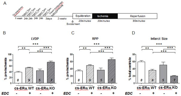
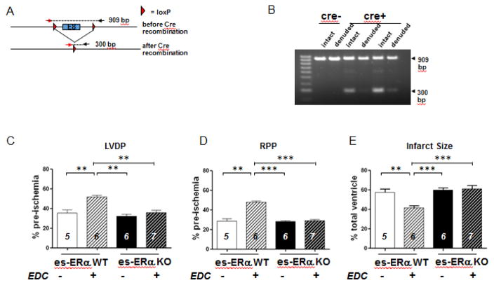
Steroid hormone receptors including estrogen receptors (ER) classically function as ligand-regulated transcription factors. However, estrogens also elicit cellular effects through binding to extra-nuclear ER (ERα, ERβ, and G protein-coupled ER or GPER) that are coupled to kinases. How extra-nuclear ER actions impact cardiac ischemia-reperfusion (I/R) injury is unknown. We treated ovariectomized wild-type female mice with estradiol or an estrogen-dendrimer conjugate (EDC), which selectively activates extra-nuclear ER, or vehicle interventions for two weeks. I/R injury was then evaluated in isolated Langendorff perfused hearts. Two weeks of treatment with estradiol significantly decreased infarct size and improved post-ischemic contractile function. Similarly, EDC treatment significantly decreased infarct size and increased post-ischemic functional recovery compared to vehicle-treated hearts. EDC also caused an increase in myocardial protein S-nitrosylation, consistent with previous studies showing a role for this post-translational modification in cardioprotection. In further support of a role for S-nitrosylation, inhibition of nitric oxide synthase, but not soluble guanylyl cyclase blocked the EDC mediated protection. The administration of ICI182,780, which is an agonist of G-protein coupled estrogen receptor (GPER) and an antagonist of ERα and ERβ, did not result in protection; however, ICI182,780 significantly blocked EDC-mediated cardioprotection, indicating participation of ERα and/or ERβ. In studies determining the specific ER subtype and cellular target involved, EDC decreased infarct size and improved functional recovery in mice lacking ERα in cardiomyocytes. In contrast, protection was lost in mice deficient in endothelial cell ERα. Thus, extra-nuclear ERα activation in endothelium reduces cardiac I/R injury in mice, and this likely entails increased protein S-nitrosylation. Since EDC does not stimulate uterine growth, in the clinical setting EDC-like compounds may provide myocardial protection without undesired uterotrophic and cancer-promoting effects.

Keywords: Cardiomyocyte; Endothelium; Estrogen receptor; Nitric oxide signaling.


Conflict of interest statement

Conflict of Interest: None declared.

Figures

Figure 1
Figure 1
Two-week treatment with estrogen and EDC improves functional recovery and infarct size. (A) I/R protocol following subcutaneous Alzet minipumps implantation in C57BL/6J. Mice were bilaterally ovariectomized at 10 weeks of age. After two weeks of recovery from the surgery, minipumps were implanted for drug treatments for two weeks. The hearts were then excised and cannulated to a Langendorff perfusion apparatus. For all the treatment groups, hearts were equilibrated for 20 minutes, subjected to 30 minutes of ischemia and reperfused for 90 minutes. (B–C) Recovery following two weeks of treatment with estradiol. Recoveries of heart functional parameters (LVDP and RPP) post-ischemia after two weeks of estradiol treatment were expressed as percentage of their pre-ischemic values. (D) To determine infarct size, hearts were perfused with 1% 2,3,5-triphenyltetrazoliumchloride (TTC) following reperfusion and ImageJ software was used to analyze the infarct size, which was expressed as the percentage of total area of cross-sectional slices (n=6 for each group, **, p<0.01). (E–F) Recovery following two weeks of treatment with EDC. Recoveries of heart functional parameters (LVDP and RPP) post-ischemia after two weeks of EDC treatment were expressed as percentage of their pre-ischemic values. (G) Infarct size was determined as described above (n=6 for each group, **, p<0.01).
Figure 2
Figure 2. Pharmacological inhibition of nitric oxide synthase or ERα/ERβ blocks the improved functional recovery by EDC treatment in ovariectomized wild type mice
(A–B) Post-ischemic recoveries of heart functional parameters (LVDP and RPP) in ovariectomized mice treated with L-NAME (a selective inhibitor of nitric oxide synthase), ODQ (an inhibitor of soluble guanylyl cyclase) or ICI182,780 (a selective inhibitor of ERα and ERβ and an activator of GPER were expressed as percentage of their pre-ischemic values. (C) Infarct size was determines as described in Figure 1D. Numbers inside each bar represent the number of mice used for each group, ***, p<0.0001.
Figure 3
Figure 3
Two-week EDC treatment leads to protein S-nitrosation changes identified by 2D CyDye-maleimide DIGE. Representative 2D CyDye-maleimide DIGE gel electrophoresis from three independent experiments. (A) Dendrimer treated group were labeled with Cy3-malemide (green) and (B) EDC treated group with Cy5-maleimide (red). (C) Overlay of S-nitrosation in EDC or vehicle treated heart.
Figure 4
Figure 4. Cardiomyocyte-specific ERα knock-out mice generation
(A) Anti-ERα Western Blot. Three samples for each group, cs-ERα WT and KO mouse heart total homogenates were loaded in the same gel and were probed with anti-ERα antibody. The amount of protein per well was checked by probing the membrane with anti-β-actin antibody. (B) Densitometry analysis show the ratio between the densitometric values of the ERα bands and those bands detected with the anti-β-actin antibody. (C–D) Post-ischemic recoveries of heart functional parameters (LVDP and RPP) in ovariectomized cs-ERα WT and KO mice treated with tamoxifen (Tam) for five days were expressed as percentage of their pre-ischemic values. (E) Infarct size was determined as described in Figure 1D. Numbers inside each bar represent the number of mice used for each group, *, p=0.0023.
Figure 5
Figure 5
Treatment for two weeks with EDC improves functional recovery and infarct size in cardiomyocyte-specific ERα knock out mice. (A) I/R protocol following subcutaneous Alzet minipumps implantation in cs-ERα KO and WT mice. Mice were bilaterally ovariectomized at 10 weeks of age. After 1 week of recovery from the ovariectomy surgery, tamoxifen was injected intraperitoneally daily for five days, and after two days minipumps were implanted for drug treatments for two weeks. (B–C) Post-ischemic recoveries of heart functional parameters (LVDP and RPP) after two weeks of EDC or dendrimer vehicle treatment were expressed as percentage of their pre-ischemic values. (D) Infarct size was determined as described in Figure 1D. Numbers inside each bar represent the number of mice used for each group, **, p<0.001; ***p<0.0001.
Figure 6
Figure 6. EDC mediated protection is blocked in mice with endothelial specific loss of ERα
Characterization of ERα expression in ERαfl/fl;VECad-Cre mice. (A–B) To evaluate effective gene excision in the endothelium, genotyping was performed on DNA isolated from intact versus endothelium-denuded aorta samples from ERαfl/fl and ERαfl/fl;VECad-Cre mice. PCR with a single primer set yielded products that were 909 bp versus 300 bp in size before versus after Cre-mediated recombination, respectively. Whereas samples from either intact or endothelium-denuded ERαfl/fl mice yielded only a 909 bp product (Lanes 1,2), samples from ERαfl/fl;VECad-Cre mice yielded both 909 bp and 300 bp products, with far less 300 bp product following endothelial denudation. (C–D) Endothelial specific ERα-KO mice were bilaterally ovariectomized at 10 weeks of age. After 1 week of recovery minipumps were implanted for drug treatments for two weeks. Post-ischemic recoveries of heart functional parameters (LVDP and RPP) after two weeks of EDC or dendrimer vehicle treatment were expressed as percentage of their pre-ischemic values. (E) Infarct size was determined as described in Figure 1D. Numbers inside each bar represent the number of mice used for each group, **, p<0.001; ***p<0.0001.

Similar articles

Cited by

References

    1. Mendelsohn ME, Karas RH. Molecular and cellular basis of cardiovascular gender differences. Science. 2005;308:1583–7. - PubMed
    1. Barrett-Connor E, Bush TL. Estrogen and coronary heart disease in women. Jama. 1991;265:1861–7. - PubMed
    1. Hodis HN, Mack WJ, Azen SP, Lobo RA, Shoupe D, Mahrer PR, et al. Hormone therapy and the progression of coronary-artery atherosclerosis in postmenopausal women. The New England journal of medicine. 2003;349:535–45. - PubMed
    1. Grady D, Herrington D, Bittner V, Blumenthal R, Davidson M, Hlatky M, et al. Cardiovascular disease outcomes during 6.8 years of hormone therapy: Heart and Estrogen/progestin Replacement Study follow-up (HERS II) Jama. 2002;288:49–57. - PubMed
    1. Manson JE, Chlebowski RT, Stefanick ML, Aragaki AK, Rossouw JE, Prentice RL, et al. Menopausal hormone therapy and health outcomes during the intervention and extended poststopping phases of the Women’s Health Initiative randomized trials. JAMA. 2013;310:1353–68. - PMC - PubMed

MeSH terms

LinkOut - more resources