. 2017 Aug:12:872-882.
doi: 10.1016/j.redox.2017.04.023. Epub 2017 Apr 19.

Catalytic oxidant scavenging by selenium-containing compounds: Reduction of selenoxides and N-chloramines by thiols and redox enzymes

Affiliations

Catalytic oxidant scavenging by selenium-containing compounds: Reduction of selenoxides and N-chloramines by thiols and redox enzymes

Luke Carroll et al. Redox Biol. 2017 Aug.

Abstract

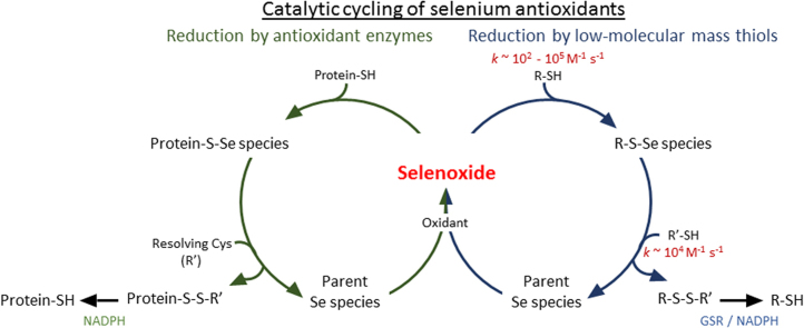
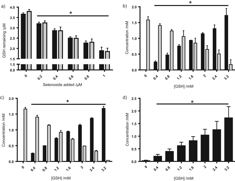
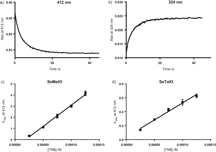
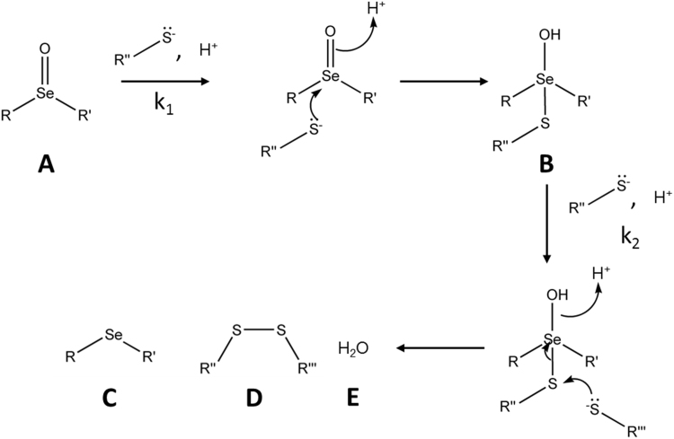
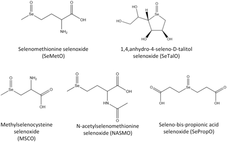
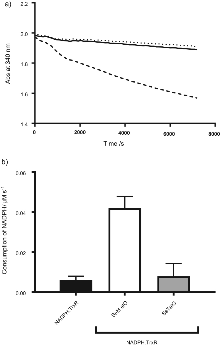
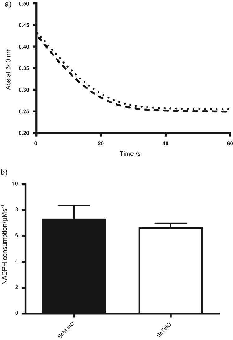
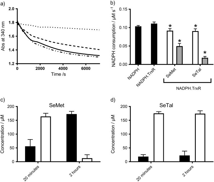
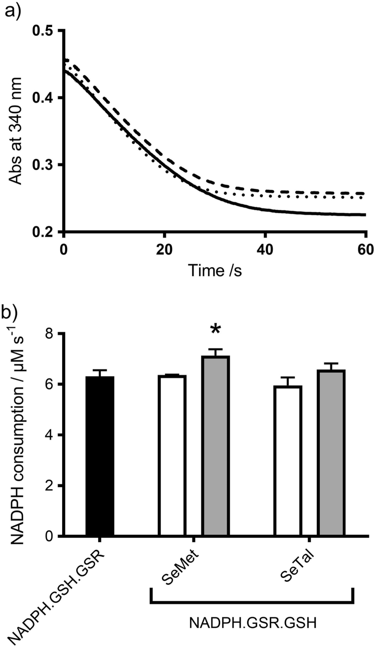
Myeloperoxidase produces strong oxidants during the immune response to destroy invading pathogens. However, these oxidants can also cause tissue damage, which contributes to the development of numerous inflammatory diseases. Selenium containing compounds, including selenomethionine (SeMet) and 1,4-anhydro-5-seleno-D-talitol (SeTal), react rapidly with different MPO-derived oxidants to form the respective selenoxides (SeMetO and SeTalO). This study investigates the susceptibility of these selenoxides to undergo reduction back to the parent compounds by intracellular reducing systems, including glutathione (GSH) and the glutathione reductase and thioredoxin reductase systems. GSH is shown to reduce SeMetO and SeTalO, with consequent formation of GSSG with apparent second order rate constants, k2, in the range 103-104M-1s-1. Glutathione reductase reduces both SeMetO and SeTalO at the expense of NADPH via formation of GSSG, whereas thioredoxin reductase acts only on SeMetO. The presence of SeMet and SeTal also increased the rate at which NADPH was consumed by the glutathione reductase system in the presence of N-chloramines. In contrast, the presence of SeMet and SeTal reduced the rate of NADPH consumption by the thioredoxin reductase system after addition of N-chloramines, consistent with the rapid formation of selenoxides, but only slow reduction by thioredoxin reductase. These results support a potential role of seleno compounds to act as catalytic scavengers of MPO-derived oxidants, particularly in the presence of glutathione reductase and NADPH, assuming that sufficient plasma levels of the parent selenoether can be achieved in vivo following supplementation.

Keywords: Antioxidants; Glutathione; Glutathione reductase; Hypochlorous acid; Myeloperoxidase; N-chloramines; Selenium; Thioredoxin reductase.


Figures

fx1
Graphical abstract
Fig. 1
Fig. 1
Selenoxides formed on SeMet and SeTal consume GSH to produce GSSG and the parent SeMet or SeTal. (a) SeMetO (black bars) or SeTalO (grey bars) (0–1 µM) was added to GSH (4 µM) and incubated for 10 min before remaining thiol was assessed by the ThioGlo assay. (b–d) GSH (0–3.2 mM) was added to (b,d) SeMetO (1.6 mM) or (c) SeTalO (1.6 mM) and incubated for 10 min before analysis by HPLC and determination of concentrations of (b) SeMetO (grey bars) and SeMet (black bars), (c) SeTalO (grey bars) and SeTal (black bars), and (d) GSSG. Data represent mean±SD from 3 independent experiments. * indicates significant difference (p<0.05) from the corresponding control where no selenoxide or GSH were added, by one-way ANOVA with Tukey's post-hoc test.
Fig. 2
Fig. 2
Kinetic analysis of the reduction of SeMetO by TNB. (a,b) Stopped flow kinetic traces measured for SeMetO (5 µM) and TNB (25 µM) in phosphate buffer (pH 7.4, 0.1 M) using a 2 mm path length cell at 22 °C measured at (a) 412 and (b) 324 nm. A loss in absorbance was observed at 412 nm, indicating TNB consumption, with a concomitant increase in absorbance at 324 nm, indicating DTNB formation. (c,d) The observed rate constants, kobs, were determined from the kinetic plots and kobs was plotted against the initial TNB concentration for (c) SeMetO or (d) SeTalO. Data represent mean±SD of three independent experiments.
Scheme 1
Scheme 1
Proposed two-step mechanism of selenoxide reduction by thiols. Initial reaction of selenoxides (A) with thiols is proposed to lead to the formation of a seleno-sulfide intermediate (B). The rate constant for the initial reaction is designated as k1. A second thiol then reacts with the intermediate with a rate constant k2, producing a selenoether (C), disulfide (D) and water (E) as products.
Fig. 3
Fig. 3
Structures of selenoxides used in the determination of the mechanisms and kinetics of selenoxide reduction by thiols.
Fig. 4
Fig. 4
Stopped flow kinetic data for the reduction of SeTalO and SeMetO by GSH. (a,b) SeTalO (125 µM) was mixed with GSH (0.5 mM) in phosphate buffer (pH 7.4, 0.1 M) at 22 °C using a 10 mm path length cell. (a) shows the absorbance changes over 0.75 s monitored at wavelengths between 240 and 300 nm and (b) shows the absorbance change monitored at 270 nm. (c,d) SeMetO (125 µM) was mixed with GSH (0.5 mM) in phosphate buffer (pH 7.4, 0.1 M) at 22 °C. (c) Shows the absorbance changes over 0.2 s monitored at wavelengths between 240 and 300 nm and (d) shows the absorbance change monitored at 240 nm where two-phase kinetics are observed. Data represent three independent experiments.
Fig. 5
Fig. 5
Enzymatic reduction of SeMetO and SeTalO by the TrxR system. Panel (a) shows typical absorbance changes of NADPH at 340 nm over time with SeMetO (200 µM; dashed line), or SeTalO (200 µM; dotted line) on addition to TrxR (25 nM) and NADPH (700 µM). The absorbance change of the assay mixture in the absence of selenoxide is shown by the solid line. Panel (b) shows the rate of consumption of NADPH by fitting a straight line to the first 10 min of the data represented in (a). For panel (b), data represent mean±SD from 3 independent experiments.
Fig. 6
Fig. 6
Enzymatic reduction of SeMetO and SeTalO by the GSR system. Panel (a) shows typical absorbance changes of NADPH at 340 nm over time with SeMetO (200 µM; dashed line), or SeTalO (200 µM; dotted line) on addition to GSR (25 nM), GSH (400 µM) and NADPH (500 µM) using a stopped-flow system with 2 mm path length cell. Panel (b) shows the initial rate of absorbance decrease over the first 10 s of data represented in (a). For panel (b), data represent mean±SD from 3 independent experiments.
Fig. 7
Fig. 7
Reduction of N-chloramines by the TrxR system in the presence of SeMet or SeTal. Panel (a) shows typical absorbance decrease of NADPH observed at 340 nm over time on addition of TauCl (200 µM) to NADPH (700 µM; solid line) in the presence of TrxR (25 nM; dot-dashed line) and SeMet (200 µM; dashed line) or SeTal (200 µM; dotted line). Panel (b) shows the rate of consumption of NADPH measured by fitting a straight line to the first 25 min of the data presented in (a), with data from experiments performed with 20 µM (white bars) and 200 µM (grey bars) SeMet and SeTal. Panel (c) shows the recovery of SeMet (black bars) from SeMetO (white bars) as determined by HPLC after TauCl was added to TrxR/NADPH in the presence of 200 µM SeMet. Panel (d) is as (c) but with SeTal. For panels (b)–(d), data represent mean±SD from 3 independent experiments. * Indicates significant difference (p<0.05) from the control by one-way ANOVA with Tukey's post-hoc test.
Fig. 8
Fig. 8
Reduction of chloramines by the GSR system in the presence of SeMet or SeTal. Panel (a) shows the typical decrease of NADPH measured at 340 nm over time on addition of TauCl (200 µM; solid line), to GSR (25 nM), GSH (400 µM) and NADPH (500 µM) in the presence of SeMet (200 µM; dashed line) or SeTal (200 µM; dotted line) using a stopped-flow system with 2 mm path length cell. Panel (b) shows the initial rate of absorbance decrease over the first 10 s of data represented in (a), with data from experiments performed with 20 µM (white bars) and 200 µM (grey bars) SeMet and SeTal. For panel (b), data represent mean±SD from 3 independent experiments. * Indicates significant difference (p<0.05) from the control by one-way ANOVA with Tukey's post-hoc test.

References

    1. Davies M.J., Hawkins C.L., Pattison D.I., Rees M.D. Mammalian heme peroxidases: from molecular mechanisms to health implications. Antioxid. Redox Signal. 2008;10:1199–1234. - PubMed
    1. Hampton M.B., Kettle A.J., Winterbourn C.C. Inside the neutrophil phagosome: oxidants, myeloperoxidase, and bacterial killing. Blood. 1998;92:3007–3017. - PubMed
    1. Rayner B.S., Love D.T., Hawkins C.L. Comparative reactivity of myeloperoxidase-derived oxidants with mammalian cells. Free Radic. Biol. Med. 2014;71:240–255. - PubMed
    1. Thomas E.L., Grisham M.B., Jefferson M.M. Preparation and characterization of chloramines. Methods Enzymol. 1986;132:569–585. - PubMed
    1. Klebanoff S.J. Myeloperoxidase: friend and foe. J. Leukoc. Biol. 2005;77:598–625. - PubMed

Publication types