. 2017 Jun 16;292(24):10239-10249.
doi: 10.1074/jbc.M117.785022. Epub 2017 Apr 30.

Lysine desuccinylase SIRT5 binds to cardiolipin and regulates the electron transport chain

Affiliations

Lysine desuccinylase SIRT5 binds to cardiolipin and regulates the electron transport chain

Yuxun Zhang et al. J Biol Chem. .

Abstract

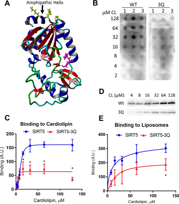
SIRT5 is a lysine desuccinylase known to regulate mitochondrial fatty acid oxidation and the urea cycle. Here, SIRT5 was observed to bind to cardiolipin via an amphipathic helix on its N terminus. In vitro, succinyl-CoA was used to succinylate liver mitochondrial membrane proteins. SIRT5 largely reversed the succinyl-CoA-driven lysine succinylation. Quantitative mass spectrometry of SIRT5-treated membrane proteins pointed to the electron transport chain, particularly Complex I, as being highly targeted for desuccinylation by SIRT5. Correspondingly, SIRT5-/- HEK293 cells showed defects in both Complex I- and Complex II-driven respiration. In mouse liver, SIRT5 expression was observed to localize strictly to the periportal hepatocytes. However, homogenates prepared from whole SIRT5-/- liver did show reduced Complex II-driven respiration. The enzymatic activities of Complex II and ATP synthase were also significantly reduced. Three-dimensional modeling of Complex II suggested that several SIRT5-targeted lysine residues lie at the protein-lipid interface of succinate dehydrogenase subunit B. We postulate that succinylation at these sites may disrupt Complex II subunit-subunit interactions and electron transfer. Lastly, SIRT5-/- mice, like humans with Complex II deficiency, were found to have mild lactic acidosis. Our findings suggest that SIRT5 is targeted to protein complexes on the inner mitochondrial membrane via affinity for cardiolipin to promote respiratory chain function.

Keywords: cardiolipin; electron transport system (ETS); mitochondria; protein acylation; sirtuin.


Conflict of interest statement

The authors declare that they have no conflicts of interest with the contents of this article

Figures

Figure 1.
Figure 1.
SIRT5 binds to cardiolipin on the inner mitochondrial membrane. A, liver mitochondria from wild-type (WT) and SIRT5−/− (KO) mice were fractionated into membrane and matrix fractions and Western blotted (25 μg of protein/lane) with anti-succinyllysine (Suc-Lys) antibody. Bottom, markers of the mitochondrial inner membrane (Tim23) and the matrix (HSP60). B, liver mitochondria from three wild-type mice were fractionated and Western blotted for SIRT5 (left) or the matrix marker protein IVD (right). Densitometry was used to measure the relative intensities of the SIRT5 bands in each fraction. C, recombinant SIRT5 and SIRT3 (200 ng) were incubated with mixed-composition unilamellar liposomes and pelleted. The pellet was washed once, and the resulting three fractions (membrane (Memb), wash, and supernatant (Sup)) were Western blotted to visualize SIRT3 and SIRT5. D, increasing amounts of cardiolipin (CL), phosphatidylethanolamine (PE), and phosphatidylcholine (PC) were spotted onto nitrocellulose, which was then incubated with 0.5 μg/ml recombinant SIRT5 diluted in blocking buffer. Bound SIRT5 was visualized by Western blotting. Avg, average.
Figure 2.
Figure 2.
An N-terminal amphipathic helix mediates SIRT5 binding to cardiolipin. A, three-dimensional molecular modeling shows an amphipathic helix at the extreme N terminus of SIRT5 (black arrow). B and C, the three charged residues on the N-terminal amphipathic helix of SIRT5 were substituted with uncharged Gln (3Q). Wild-type and mutant SIRT5 were tested for binding to a fat blot (0.5 μg of protein/ml); the resulting blot was quantified by densitometry (C). D and E, SIRT5-3Q also shows reduced binding in a pulldown assay in which 200 ng of each recombinant protein was incubated with an increasing concentration of cardiolipin (CL) liposomes, pelleted by centrifugation, and detected by Western blotting. *, p < 0.05 versus wild-type protein. Graphs in C and E represent means, and error bars represent S.D. A.U., arbitrary units.
Figure 3.
Figure 3.
SIRT5 counteracts succinylation of mitochondrial membrane proteins. A, a freshly isolated lobe of SIRT5−/− liver was stained with X-Gal to reveal the pattern of β-galactosidase expression from the SIRT5 mutant allele (blue color indicates zones expressing SIRT5). The “honeycomb” structure of the hepatic lobules can be seen outlined by the staining for β-galactosidase, which localizes to the periportal (PP) zone. The central vein (CV) can be seen as a brown spot in the middle of each lobule (red arrow as example), and no blue X-Gal staining appears in the region around the central vein. B, washed mitochondrial membranes from SIRT5−/− liver were treated with 0.5 mm succinyl-CoA (Succ-CoA) from 0 to 5 h, and the effects were visualized by Western blotting with anti-succinyllysine (Suc-Lys) antibody with Ponceau S staining as a loading control. Below the Ponceau image are the -fold changes in global succinylation estimated by densitometry. C, quadruplicate samples of succinyl-CoA-treated SIRT5−/− membranes were treated with either inactive mutant recombinant SIRT5 H158Y (Control) or wild-type recombinant SIRT5 (SIRT5-Tx), and the resulting desuccinylation was visualized by Western blotting with anti-succinyllysine antibody. D, the blot from C was quantified by densitometry of the entire Western blot lanes; shown are the means of the four Control and four SIRT5-Tx lanes; error bars represent S.D. E and F, the remainder of the eight samples shown in C were subjected to quantitative mass spectrometry to identify the specific lysine residues targeted by SIRT5 (see supplemental Table 1). Shown is a summary of the unique succinylation sites identified and the number of SIRT5 target sites defined as showing a statistically significant change at p < 0.05. F is a volcano plot (log p value plotted against log -fold change) of the mass spectrometry results.
Figure 4.
Figure 4.
SIRT5 targets the mitochondrial respiratory chain. A, the top SIRT5-targeted pathways as identified by Reactome pathway analysis. The top pathway, electron transport, was selected for further analysis. B, a total of 223 succinylated lysine residues on the ETC showed significant change upon SIRT5 treatment. Shown are the -fold changes for sites identified on each of the five ETC complexes with the total number of sites per complex shown in parentheses below the x axis. The red lines indicate the average -fold change for each complex. C, validation that ETC Complex II SDHA is succinylated in vivo using anti-SDHA immunoprecipitation from mouse liver extracts followed by Western blotting with anti-succinyllysine (Su-K) antibody. Shown below the blot are the -fold changes in succinylation between the fed and fasted mice. This experiment was repeated once with similar results. D and E, mitochondrial respiration was assessed in a human SIRT5-deficient HEK293 cell line in either the fed state (normal DMEM) or after 3 h of glucose starvation (0.1 mm glucose). Cells were permeabilized with digitonin and probed in an Oroboros Oxygraph-2K for pyruvate/malate-driven respiration (primarily Complex I) or succinate-driven respiration (Complex II). Within each experiment, respiration rates were normalized to the maximal respiration observed after addition of a chemical uncoupler (CCCP). Shown are the means of four separate experiments; error bars represent S.D. *, p < 0.05. CI–CV, Complexes I–V.
Figure 5.
Figure 5.
SIRT5 deficiency does not affect the abundance of the respiratory chain complexes in liver mitochondria. A, blue native gel electrophoresis of wild-type (WT) and SIRT5−/− (KO) liver mitochondria. Respiratory chain Complexes I, III, IV, and V (ATP synthase) and supercomplexes (SC) are indicated with arrows. B, Western blot of the two soluble subunits of Complex II (CII) (SDHA and SDHB) in liver mitochondrial extracts with Tim23 as a loading control. Densitometry was used to evaluate the protein levels in each lane relative to Tim23, and the results are shown in the bar graph (error bars represent S.D.).
Figure 6.
Figure 6.
Complex II and ATP synthase activities are reduced in SIRT5−/− mouse liver. A and B, an Oroboros Oxygraph-2K was used to measure pyruvate/malate (A) and succinate (B) respiration in liver homogenates normalized to maximal respiration induced by the chemical uncoupler CCCP. C and D, an immunocapture method was used to measure Complex I (C) and Complex II (D) enzyme activities. E, in-gel activity stains for Complexes III (CIII) and IV (CIV) performed after blue native gel electrophoresis of digitonin-permeabilized mitochondria. Densitometry was used to generate the bar graphs shown below the stained gel. F, complex V (ATP synthase) ATPase activity in liver mitochondrial lysates. Bars represent means, and error bars represent S.D. with n = 6 mice for A and B; n = 8 mice for C, D, and F, and n = 4 mice for E. *, p < 0.01; **, p < 0.001 by t test.
Figure 7.
Figure 7.
SIRT5 targets lysines at the protein-lipid interface of Complex II. A, details regarding six lysine residues on Complex II subunit SDHB (identified through quantitative mass spectrometry) that are predicted to be at the protein-lipid interface. All showed statistically significant reductions in succinylation following treatment with SIRT5 (SIRT5 Tx) (n = 4). B, three-dimensional model created from crystal structure of porcine Complex II illustrating the location of the six residues from A. Note that some of the residues are oriented into the page and cannot be seen. The ubiquinone-binding site is indicated with an arrow, and iron-sulfur clusters are rendered in magenta. C, blood lactate levels from unstressed SIRT5−/− mice (left) and FAO-deficient LCAD−/− mice (right). The SIRT5 graph shows means for n = 8 mice (**, p < 0.001), and the LCAD graph shows means for n = 6 mice; all error bars represent S.D.

References

    1. Zhang Y., Bharathi S. S., Rardin M. J., Uppala R., Verdin E., Gibson B. W., and Goetzman E. S. (2015) SIRT3 and SIRT5 regulate the enzyme activity and cardiolipin binding of very long-chain acyl-CoA dehydrogenase. PLoS One 10, e0122297. - PMC - PubMed
    1. Fould B., Garlatti V., Neumann E., Fenel D., Gaboriaud C., and Arlaud G. J. (2010) Structural and functional characterization of the recombinant human mitochondrial trifunctional protein. Biochemistry 49, 8608–8617 - PubMed
    1. Kashfi K., Mynatt R. L., Park E. A., and Cook G. A. (2011) Membrane microenvironment regulation of carnitine palmitoyltranferases I and II. Biochem. Soc. Trans. 39, 833–837 - PubMed
    1. Mileykovskaya E., and Dowhan W. (2014) Cardiolipin-dependent formation of mitochondrial respiratory supercomplexes. Chem. Phys. Lipids 179, 42–48 - PMC - PubMed
    1. Wang Y., Mohsen A. W., Mihalik S. J., Goetzman E. S., and Vockley J. (2010) Evidence for physical association of mitochondrial fatty acid oxidation and oxidative phosphorylation complexes. J. Biol. Chem. 285, 29834–29841 - PMC - PubMed

MeSH terms

LinkOut - more resources