

Thrombosis in an Internal Jugular Vein and an Upper Limb Deep Vein Treated with Edoxaban
- PMID: 28458311
- PMCID: PMC5478566
- DOI: 10.2169/internalmedicine.56.7405
Thrombosis in an Internal Jugular Vein and an Upper Limb Deep Vein Treated with Edoxaban
Abstract
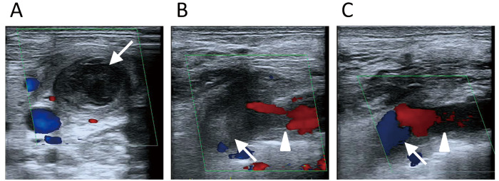
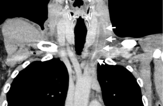
A 45-year-old man complained of swelling of the left side of his neck and left upper limb. Ultrasonography and enhanced computed tomography (CT) revealed thrombosis of the left internal jugular, subclavian, and brachiocephalic vein. Based on various examinations, the patient was diagnosed with idiopathic venous thrombosis early in his clinical course. There were no findings to suggest malignancy or abnormal coagulability. However, two months after the start of treatment, the patient was diagnosed with gastric cancer. Despite the presence of Trousseau syndrome, treatment with edoxaban (an oral anticoagulant), reduced the swelling dramatically without any bleeding complications.
Keywords: Trousseau syndrome; oral anticoagulant; upper-extremity deep vein thrombosis.
Figures
Similar articles
-
Subclavian vein thrombosis with internal jugular vein extension in an Australian rules football player.Med J Aust. 2018 Oct 15;209(8):335-336. doi: 10.5694/mja18.00335. Med J Aust. 2018. PMID: 30309306 No abstract available.
-
Edoxaban was Effective for Treating Renal Vein Thrombosis in a Patient with Nephrotic Syndrome.Intern Med. 2017 Sep 1;56(17):2307-2310. doi: 10.2169/internalmedicine.8742-16. Epub 2017 Aug 10. Intern Med. 2017. PMID: 28794382 Free PMC article.
-
Alternative venous outflow by brachial to jugular vein vascular access for hemodialysis in the exhausted upper extremities.J Vasc Access. 2015 Jul-Aug;16(4):269-74. doi: 10.5301/jva.5000363. Epub 2015 Feb 4. J Vasc Access. 2015. PMID: 25656257
-
Edoxaban: front-line treatment for brachiocephalic vein thrombosis in primitive mediastinal seminoma: A case report and literature review.Medicine (Baltimore). 2022 Aug 26;101(34):e29429. doi: 10.1097/MD.0000000000029429. Medicine (Baltimore). 2022. PMID: 36042679 Free PMC article. Review.
-
[Ovarian hyperstimulation syndrome and thrombosis. Apropos of a case of thrombosis of the internal jugular vein. Review of the literature].J Gynecol Obstet Biol Reprod (Paris). 1994;23(7):778-83. J Gynecol Obstet Biol Reprod (Paris). 1994. PMID: 7822710 Review. French.
Cited by
-
MiR-411 suppressed vein wall fibrosis by downregulating MMP-2 via targeting HIF-1α.J Thromb Thrombolysis. 2018 Feb;45(2):264-273. doi: 10.1007/s11239-017-1596-8. J Thromb Thrombolysis. 2018. PMID: 29264695
-
Internal Jugular and Subclavian Vein Thrombosis in a Post-liver Transplant Patient.Cureus. 2020 Jan 3;12(1):e6557. doi: 10.7759/cureus.6557. Cureus. 2020. PMID: 31942270 Free PMC article.
-
Unprovoked internal jugular vein thrombosis: a case report and literature review.Thromb J. 2021 Jan 6;19(1):2. doi: 10.1186/s12959-020-00246-7. Thromb J. 2021. PMID: 33407545 Free PMC article.
-
Internal Jugular Vein Thrombosis: Etiology, Symptomatology, Diagnosis and Current Treatment.Diagnostics (Basel). 2021 Feb 23;11(2):378. doi: 10.3390/diagnostics11020378. Diagnostics (Basel). 2021. PMID: 33672254 Free PMC article. Review.
-
Idiopathic Internal Jugular Vein and Subclavian Vein Thrombosis: A Rare Case Report.Cureus. 2019 Feb 4;11(2):e4005. doi: 10.7759/cureus.4005. Cureus. 2019. PMID: 31001459 Free PMC article.
References
-
- Sajid MS, Ahmed N, Desai M, Baker D, Hamilton G. Upper limb deep vein thrombosis: a literature review to streamline the protocol for management. Acta Haematol 118: 10-18, 2007. - PubMed
-
- Verso M, Agnelli G. Venous thromboembolism associated with long term use of central venous catheters in cancer patients. J Clin Oncol 19: 3665-3675, 2003. - PubMed
-
- Lindbland B, Tengborn L, Bergqvist D. Deep vein thrombosis of the axillary and subclavian veins: epidemiological data, effects of different types of treatment and late sequelae. Eur J Vasc Surg 2: 161-165, 1988. - PubMed
-
- Shameem M, Akhtar J, Bhargava R, Ahmed Z, Baneen U, Khan NA. Internal jugular vein thrombosis: a rare presentation of mediastinal lymphoma. Respir Med CME 3: 273-275, 2010.
Publication types
MeSH terms
Substances
LinkOut - more resources
Full Text Sources
Other Literature Sources
Medical