

Rhinocerebral Mucormycosis: Report of a Rare Case
- PMID: 28458494
- PMCID: PMC5390232
- DOI: 10.4314/ejhs.v27i1.11
Rhinocerebral Mucormycosis: Report of a Rare Case
Abstract
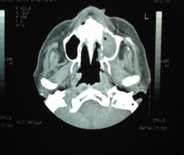
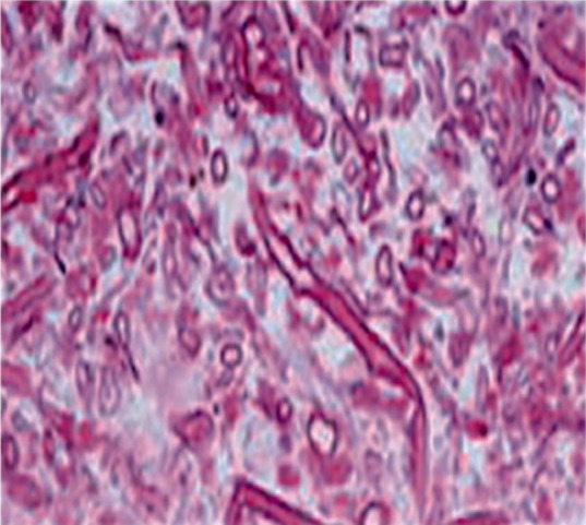
Background: Mucormycosis is one of the rapidly progressing and lethal form of fungal infection which involves the nose and paranasal sinuses of the head and the neck regions. Mucormycosis also remains a threat to patients with uncontrolled diabetes or other predisposing systemic conditions. It manifests as rhinocerebral, pulmonary, gastrointestinal, cutaneous or disseminated form. The underlying conditions can influence clinical presentation and often delay diagnosis, with resultant poor outcomes.
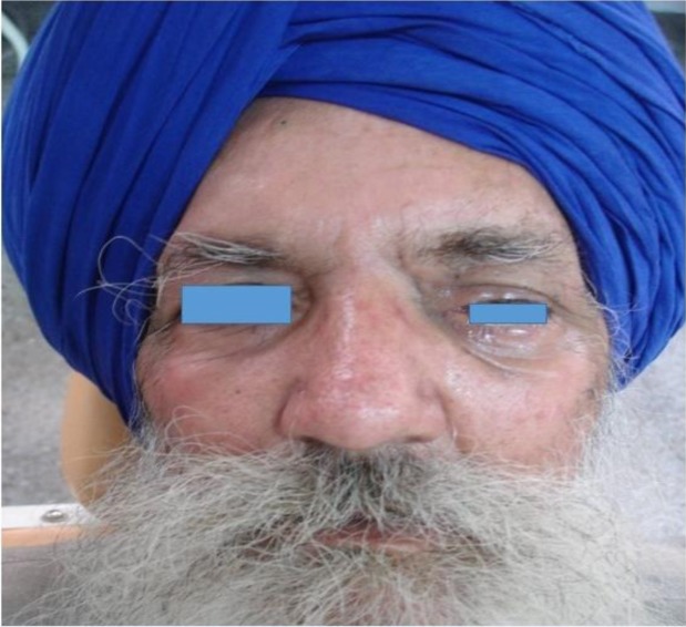
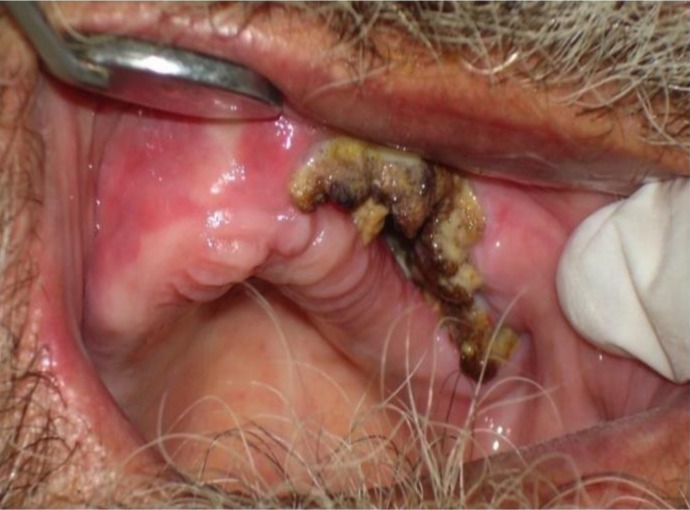
Case details: We report a case of rhinocerebral mucormycosis in a 75 year-old diabetic patient with emphasise on diagnosis, treatment and survival options of patient from this potentially fatal fungal infection. Extra oral examination revealed mild non-tender swelling on the face, unable to see from left eye, impaired sense of smell, difficulty in speech and nasal stuffiness. Intra-oral examination showed necrosis of mucosa and underlying bone in relation to canine to the tuberosity area of the left vestibular region of the maxilla.
Conclusion: Timely diagnosis is critical to survival and minimization of morbidity. Institution of surgical and medical therapy is critical in maximizing the likelihood of good outcome.
Keywords: Mucormycosis; fungal; maxillary sinus; systemic.
Conflict of interest statement
Competing Interests: The authors declare that this manuscript was approved by all authors in its form and that no competing interest exists.
Figures
References
-
- Dimaka K, Mallis A, Naxakis SS, Marangos M, Papadas TA, Stathas T, et al. Chronic rhinocerebral mucormycosis: a rare case report and review of the literature. Mycoses. 2014;57:699–702. - PubMed
-
- Margo CE, Linden C, Strickland-Marmol LB, Denietolis AL, McCaffrey JC, Kirk N. Rhinocerebral mucormycosis with perineural spread. Ophthal Plast Reconstr Surg. 2007;23:326–327. - PubMed
-
- Berdai MA, Labib S, Harandou M. Rhinocerebral mucormycosis complicating ketoacidosis diabetes. Presse Med. 2016;45:145–146. - PubMed
-
- Reddy SS, Rakesh N, Chauhan P, Sharma S. Rhinocerebral Mucormycosis Among Diabetic Patients: An Emerging Trend. Mycopathologia. 2015;180:389–396. - PubMed
Publication types
MeSH terms
Substances
LinkOut - more resources
Full Text Sources