Review
. 2017 May-Jun;18(3):413-426.
doi: 10.3348/kjr.2017.18.3.413. Epub 2017 Apr 3.

Hepatic Lesions that Mimic Metastasis on Radiological Imaging during Chemotherapy for Gastrointestinal Malignancy: Recent Updates

Affiliations
Review

Hepatic Lesions that Mimic Metastasis on Radiological Imaging during Chemotherapy for Gastrointestinal Malignancy: Recent Updates

Sung-Hye You et al. Korean J Radiol. 2017 May-Jun.

Abstract

During chemotherapy in patients with gastrointestinal malignancy, the hepatic lesions may occur as chemotherapy-induced lesions or tumor-associated lesions, with exceptions for infectious conditions and other incidentalomas. Focal hepatic lesions arising from chemotherapy-induced hepatopathies (such as chemotherapy-induced sinusoidal injury and steatosis) and tumor-associated eosinophilic abscess should be considered a mimicker of metastasis in patients with gastrointestinal malignancy. Accumulating evidence suggests that chemotherapy for gastrointestinal malignancy in the liver has roles in both the therapeutic effects for hepatic metastasis and injury to the non-tumor bearing hepatic parenchyma. In this article, we reviewed the updated concept of chemotherapy-induced hepatopathies and tumor-associated eosinophilic abscess in the liver, focusing on the pathological and radiological findings. Awareness of the causative chemo-agent, pathophysiology, and characteristic imaging findings of these mimickers is critical for accurate diagnosis and avoidance of unnecessary exposure of the patient to invasive tissue-based diagnosis and operations.

Keywords: Chemotherapy-induced focal hepatopathy; Oxaliplatin; Peliosis; Sinusoidal obstructive syndrome; Steatohepatitis; Steatosis.


Figures

Fig. 1
Fig. 1. Algorithm of pathophysiology of chemotherapy-induced hepatopathy.
GI = gastrointestinal, MMP = matrix metalloproteinase, RBCs = red blood cells, 5-FU = fluorouracil
Fig. 2
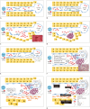
Fig. 2. Schematic illustration of pathophysiology of chemotherapy-induced sinusoidal injury.
A. Normal sinusoid. B. SEC injury and disruption of sinusoidal wall integrity. C. Peliosis with fresh blood. D. Peliosis with old blood. E. Perisinusoidal fibrosis. F. Sinusoidal dilatation. G. Nodular regenerative hyperplasia. H. Chemotherapy-induced sinusoidal injury. RBCs = red blood cells, SEC = sinusoidal endothelial cell
Fig. 3
Fig. 3. Three types of hepatic distributions of post-oxaliplatin heterogeneity of liver parenchyma on contrast-enhanced portal phase computed tomography scans.
A. Peripheral distribution: liver shows heterogeneous hypoattenuation mainly in periphery of liver. B. Multifocal distribution: liver shows multifocal hypoattenuations (arrows) in liver. C. Diffuse distribution: liver shows diffuse heterogeneous hypoattenuation.
Fig. 4
Fig. 4. Magnetic resonance images of 53-year-old woman with heterogeneous reticular pattern in non-tumor bearing parenchyma on hepatobiliary-phase imaging of liver.
Pattern was detected 3 months after initiation of chemotherapy for colon cancer. After four cycles of chemotherapy with FOLFOX, variegated reticular parenchymal hypointensity on hepatobiliary phase image had newly developed in non-tumor bearing liver parenchyma.
Fig. 5
Fig. 5. Magnetic resonance images of 41-year-old man with 1-cm chemotherapy-induced focal sinusoidal injury detected 1 month after initiation of chemotherapy (FOLFOX) for colon cancer.
A. After four cycles of chemotherapy with FOLFOX, ill-defined ovoid lesion (arrow) had newly developed in segment 2 of liver. Lesion shows intermingled hypointensity with combined variegated reticular parenchymal hypointensity on hepatobiliary phase image. B. Iso-signal intensity (arrow) on fat-suppressed respiratory-triggered heavily T2-weighted image. C. No peripheral rim enhancement (arrow) is seen on arterial phase image. D. Iso-signal intensity on high b-value diffusion-weighted image (b = 800 s/mm2, arrow). E. (Upper half) cut surface of liver shows well-demarcated lesion with diffuse hemorrhage (arrowheads) in segment 2. (Lower half) microscopically, lesion shows diffuse sinusoidal dilatation with cystically dilated blood-filled spaces indicative of peliosis (*). Note degenerated red blood cells within dilated sinusoids (hematoxylin and eosin, × 200).
Fig. 6
Fig. 6. Magnetic resonance images of 58-year-old woman with chemotherapy-induced focal steatosis mimicking metastasis, which was detected 12 month after initiation of chemotherapy (FOLFOX) for colon cancer.
A. After 12 cycles of chemotherapy, ill-defined ovoid lesion (arrow) had newly developed in segment 6 of liver. Lesion shows hypointensity without combined variegated reticular parenchymal hypointensity on hepatobiliary phase image. B. No peripheral rim enhancement (arrow) is seen on portal phase of dynamic enhancement. C, D. There is high signal nodular lesion (arrow) on in-phase and signal drop (arrow) on opposed-phase of chemical shift image, respectively. E. Microscopically, liver shows microvesicular and macrovesicular steatosis. Small fat droplets are assembled around centrally located nuclei (arrowheads) and large fat droplets replace nuclei to periphery of cell (arrows) (hematoxylin and eosin, × 200).
Fig. 7
Fig. 7. Illustration of pathophysiology of tumor-associated eosinophilic abscess in liver.
GM-CSF = granulocyte-macrophage colony stimulating factor
Fig. 8
Fig. 8. Magnetic resonance images of 55-year-old man with colon cancer and surgically confirmed eosinophilic abscesses in liver.
A. Respiratory-triggered T2-weighted turbo spin-echo image shows single hyperintense nodular lesion (arrows) in right hepatic lobe. B. On T1-weighted gradient echo magnetic resonance imaging, lesion is almost isointense. C. On arterial phase, lesion appears as rim-enhancing nodule (arrows). D. On hepatobiliary phase image, lesion is clearly seen as relatively well-defined hypointense areas (arrows), and appears larger than that observed in (B). E. Ill-demarcated grayish white lesion is noted at subcapsular area of liver (arrowheads). F. Microscopically, lesion (indicated by arrowheads in E) is composed of mixed inflammatory cell infiltrates, predominantly with eosinophils (arrows), replacing normal hepatocytes (hematoxylin and eosin, × 200).
Fig. 9
Fig. 9. Schematic representation of typical magnetic resonance imaging features of hepatic pseudometastasis during chemotherapy in patients with gastrointestinal malignancy.
A = arterial phase image, ADC = apparent diffusion coefficient, b800 = diffusion weighted image (b value = 800 s/mm2), D = delayed-phase image, HBP = hepatobiliary phase, IP = in-phase magnetic resonance image, OP = opposed-phase magnetic resonance image, P = portal-phase image, Pre = precontrast T1-weighted image, T1WI = T1-weighted image, T2WI = T2-weighted image

References

    1. Han NY, Park BJ, Sung DJ, Kim MJ, Cho SB, Lee CH, et al. Chemotherapy-induced focal hepatopathy in patients with gastrointestinal malignancy: gadoxetic acid--enhanced and diffusion-weighted MR imaging with clinical-pathologic correlation. Radiology. 2014;271:416–425. - PubMed
    1. Zhou B, Yan S, Zheng S. Peliosis hepatis: a mimicker of hepatic tumors. Surg Pract. 2013;7:124–126.
    1. Arakawa Y, Shimada M, Utsunomya T, Imura S, Morine Y, Ikemoto T, et al. Oxaliplatin-related sinusoidal obstruction syndrome mimicking metastatic liver tumors. Hepatol Res. 2013;43:685–689. - PubMed
    1. Kim MJ, Kim HJ, Yi BH, Kim HK, Yun J, Kim SH, et al. A case of peliosis hepatis mimicking metastatic lesions of increasing size. Soonchunhyang Med Sci. 2012;18:141–144.
    1. Xiong WJ, Hu LJ, Jian YC, He Y, Zhou W, Guo XL, et al. Focal peliosis hepatis in a colon cancer patient resembling metastatic liver tumor. World J Gastroenterol. 2012;18:5999–6002. - PMC - PubMed

MeSH terms