Review
. 2017:2017:6027029.
doi: 10.1155/2017/6027029. Epub 2017 Mar 26.

A Review on Real-Time 3D Ultrasound Imaging Technology

Affiliations
Review

A Review on Real-Time 3D Ultrasound Imaging Technology

Qinghua Huang et al. Biomed Res Int. 2017.

Abstract

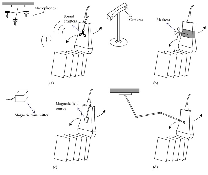
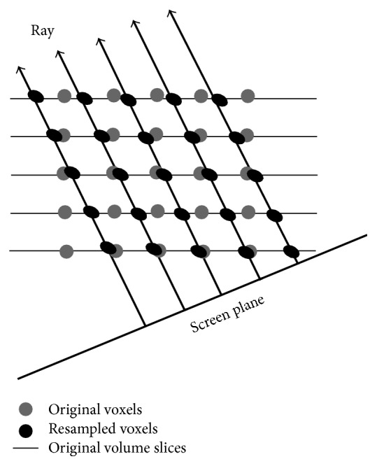
Real-time three-dimensional (3D) ultrasound (US) has attracted much more attention in medical researches because it provides interactive feedback to help clinicians acquire high-quality images as well as timely spatial information of the scanned area and hence is necessary in intraoperative ultrasound examinations. Plenty of publications have been declared to complete the real-time or near real-time visualization of 3D ultrasound using volumetric probes or the routinely used two-dimensional (2D) probes. So far, a review on how to design an interactive system with appropriate processing algorithms remains missing, resulting in the lack of systematic understanding of the relevant technology. In this article, previous and the latest work on designing a real-time or near real-time 3D ultrasound imaging system are reviewed. Specifically, the data acquisition techniques, reconstruction algorithms, volume rendering methods, and clinical applications are presented. Moreover, the advantages and disadvantages of state-of-the-art approaches are discussed in detail.


Figures

Figure 1
Figure 1
Principle of volumetric imaging with a 2D array transducer.
Figure 2
Figure 2
The consisting material of a single-unit type matrix transducer.
Figure 3
Figure 3
Schematic structure of a mechanical 3D probe.
Figure 4
Figure 4
Schematic structure of three types of mechanical scanning: (a) tilting scanning; (b) linear scanning; (c) rotational scanning.
Figure 5
Figure 5
Schematic structure of three types of position sensor: (a) acoustic sensor; (b) optimal positioner; (c) magnetic field sensor; (d) articulated arm positioner.
Figure 6
Figure 6
VNN. A normal from the voxel to two nearest frames is calculated and the nearest pixel is selected to be mapped into the voxel.
Figure 7
Figure 7
Consider the thickness of the US beam to improve inserted quality.
Figure 8
Figure 8
VBM with interpolation from the two nearest surrounding images where a normal to each image is calculated.
Figure 9
Figure 9
The probe trajectory used to find the two intersecting points on the two surrounding images is estimated.
Figure 10
Figure 10
Squared distance weighted interpolation. Pixels that fall within the spherical region make value contribution to the central voxel.
Figure 11
Figure 11
PBMs DS with a 3D ellipsoid Gaussian kernel around the pixel and the extent and weighting is determined by an ellipsoid Gaussian kernel.
Figure 12
Figure 12
PBM GFS. Gap-filling with an ellipsoid kernel around a voxel, and the PSF of the US system is used to determine the kernel shape and weighting.
Figure 13
Figure 13
Functional interpolation. The function through the input points is estimated and evaluated at regular intervals to obtain the final voxel values.
Figure 14
Figure 14
Bezier interpolation. Movement of the control window along with the sequences of B-scans for reconstruction of 3D volume.
Figure 15
Figure 15
Volume rendering using direct ray casting. Voxels along each ray are resampled via a trilinear interpolation of eight neighboring original voxels.
Figure 16
Figure 16
Fast volume rendering using shear-warp. Bilinear interpolation is used within each slice to resample each voxel along a ray from the four neighboring original voxels.
Figure 17
Figure 17
Fast volume rendering using shear-image-order. Bilinear interpolation is used within each slice to resample each voxel along a ray from the four neighboring original voxels.

References

    1. Feng X., Guo X., Huang Q. Systematic evaluation on speckle suppression methods in examination of ultrasound breast images. Applied Sciences. 2017;7(1):p. 37. doi: 10.3390/app7010037. - DOI
    1. Huang Q., Luo Y., Zhang Q. Breast ultrasound image segmentation: a survey. International Journal of Computer Assisted Radiology and Surgery. 2017;12(3):493–507. doi: 10.1007/s11548-016-1513-1. - DOI - PubMed
    1. Huang Q., Yang F., Liu L., Li X. Automatic segmentation of breast lesions for interaction in ultrasonic computer-aided diagnosis. Information Sciences. 2015;314:293–310. doi: 10.1016/j.ins.2014.08.021. - DOI
    1. Chang H., Chen Z., Huang Q., Shi J., Li X. Graph-based learning for segmentation of 3D ultrasound images. Neurocomputing. 2015;151(2):632–644. doi: 10.1016/j.neucom.2014.05.092. - DOI
    1. Welch J. N., Johnson J. A., Bax M. R., et al. Real-time freehand 3D ultrasound system for clinical applications. Medical Imaging 2001: Visualization, Display, and Image-Guided Procedures; February 2001; San Diego, Calif, USA. pp. 724–730. - DOI