

Meckel's cartilage breakdown offers clues to mammalian middle ear evolution
- PMID: 28459103
- PMCID: PMC5405799
- DOI: 10.1038/s41559-017-0093
Meckel's cartilage breakdown offers clues to mammalian middle ear evolution
Abstract
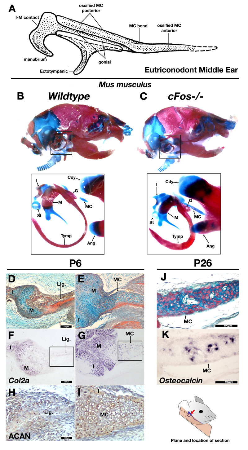
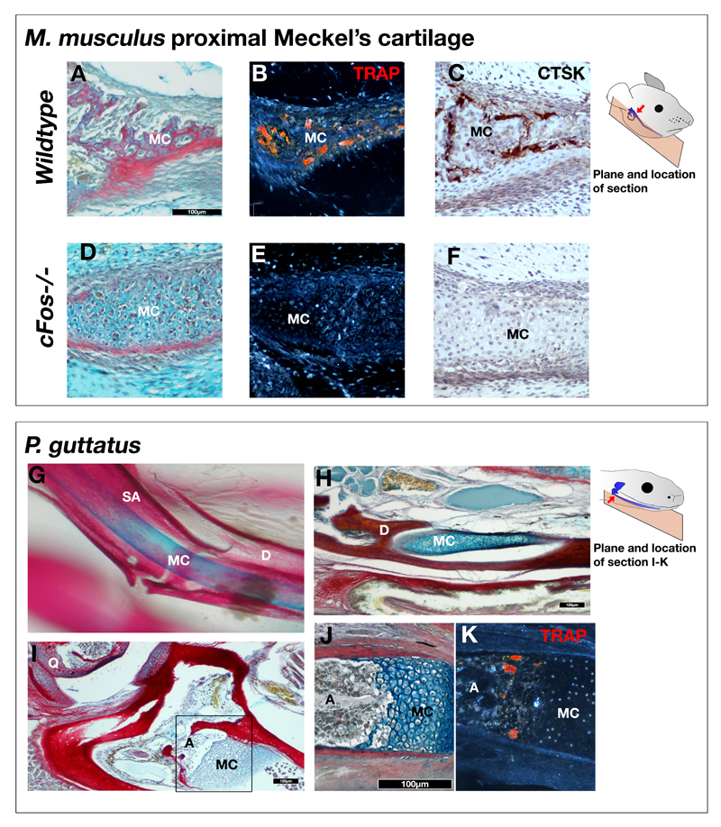
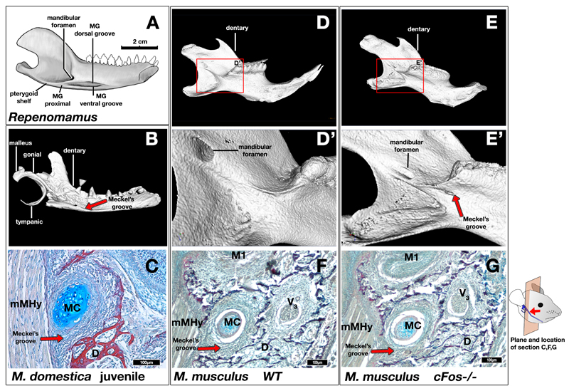
A key transformation in mammalian ear evolution was incorporation of the primary jaw joint of premammalian synapsids into the definitive mammalian middle ear of living mammals. This evolutionary transition occurred in two-steps, starting with a partial or "transitional" mammalian middle ear in which the ectotympanic and malleus were still connected to the mandible by an ossified Meckel's Cartilage (MC), as observed in many Mesozoic mammals. This was followed by MC breakdown, freeing the ectotympanic and the malleus from the mandible and creating the definitive mammalian middle ear. Here we report novel findings on the role of chondroclasts in MC breakdown, shedding light on how therian mammals lost MC connecting the ear to the jaw. Genetic or pharmacological loss of clast cells in mice and opossums leads to persistence of embryonic MC beyond juvenile stages, with MC ossification in mutant mice. The persistent MC causes a distinctive postnatal groove on the mouse dentary. This morphology phenocopies the ossified MC and Meckelian groove observed in Mesozoic mammals. Clast cell recruitment to MC is not observed in reptiles, where MC persists as a cartilaginous structure. We hypothesize that ossification of MC is an ancestral feature of mammaliaforms, and that a shift in the timing of clast cell recruitment to MC prior to its ossification is a key developmental mechanism for the evolution of the definitive mammalian middle ear in extant therians.
Conflict of interest statement
Author information The authors declare no competing financial interests.
Figures

Similar articles
-
Transitional mammalian middle ear from a new Cretaceous Jehol eutriconodont.Nature. 2011 Apr 14;472(7342):181-5. doi: 10.1038/nature09921. Nature. 2011. PMID: 21490668
-
Evolutionary development of the middle ear in Mesozoic therian mammals.Science. 2009 Oct 9;326(5950):278-81. doi: 10.1126/science.1178501. Science. 2009. PMID: 19815774
-
Mammalian development does not recapitulate suspected key transformations in the evolutionary detachment of the mammalian middle ear.Proc Biol Sci. 2016 Jan 13;283(1822):20152606. doi: 10.1098/rspb.2015.2606. Proc Biol Sci. 2016. PMID: 26763693 Free PMC article.
-
Diverse Fate of an Enigmatic Structure: 200 Years of Meckel's Cartilage.Front Cell Dev Biol. 2020 Aug 28;8:821. doi: 10.3389/fcell.2020.00821. eCollection 2020. Front Cell Dev Biol. 2020. PMID: 32984323 Free PMC article. Review.
-
Morphological evolution of the mammalian jaw adductor complex.Biol Rev Camb Philos Soc. 2017 Nov;92(4):1910-1940. doi: 10.1111/brv.12314. Epub 2016 Nov 23. Biol Rev Camb Philos Soc. 2017. PMID: 27878942 Free PMC article. Review.
Cited by
-
Making and shaping endochondral and intramembranous bones.Dev Dyn. 2021 Mar;250(3):414-449. doi: 10.1002/dvdy.278. Epub 2020 Dec 28. Dev Dyn. 2021. PMID: 33314394 Free PMC article. Review.
-
Evolution and development of the mammalian jaw joint: Making a novel structure.Evol Dev. 2023 Jan;25(1):3-14. doi: 10.1111/ede.12426. Epub 2022 Dec 11. Evol Dev. 2023. PMID: 36504442 Free PMC article. Review.
-
Middle ear innovation in Early Cretaceous eutherian mammals.Nat Commun. 2023 Oct 26;14(1):6831. doi: 10.1038/s41467-023-42606-7. Nat Commun. 2023. PMID: 37884521 Free PMC article.
-
Making sense of vertebrate senses from a neural crest and cranial placode evo-devo perspective.Trends Neurosci. 2025 Mar;48(3):213-226. doi: 10.1016/j.tins.2024.12.008. Epub 2025 Jan 23. Trends Neurosci. 2025. PMID: 39848838 Review.
-
Evolution and development of the fish jaw skeleton.Wiley Interdiscip Rev Dev Biol. 2019 Mar;8(2):e337. doi: 10.1002/wdev.337. Epub 2018 Oct 31. Wiley Interdiscip Rev Dev Biol. 2019. PMID: 30378758 Free PMC article. Review.
References
-
- Allin EF, Hopson JA. In: The Evolutionary Biology Of Hearing. Webster DB, Fay RR, Popper AN, editors. Springer-Verlag; 1992. pp. 587–614.
-
- Rowe T. Coevolution of the mammalian middle ear and neocortex. Science. 1996;273:651–654. - PubMed
-
- Luo Z-X. Transformation and diversification in early mammal evolution. Nature. 2007;450:1011–9. - PubMed
-
- Anthwal N, Tucker AS. In: From Clone To Bone: The Synergy Of Morphological And Molecular Tools In Palaeobiology. Asher RJ, Müller J, editors. Cambridge University Press; 2012. pp. 207–229.
Grants and funding
LinkOut - more resources
Full Text Sources
Other Literature Sources
