

Case Reports
doi: 10.1038/nm.4330.
Epub 2017 May 1.
Silent hippocampal seizures and spikes identified by foramen ovale electrodes in Alzheimer's disease
Affiliations
- PMID: 28459436
- PMCID: PMC5461182
- DOI: 10.1038/nm.4330
Item in Clipboard
Case Reports
Silent hippocampal seizures and spikes identified by foramen ovale electrodes in Alzheimer's disease
Nat Med.
2017 Jun.
Abstract
We directly assessed mesial temporal activity using intracranial foramen ovale electrodes in two patients with Alzheimer's disease (AD) without a history or EEG evidence of seizures. We detected clinically silent hippocampal seizures and epileptiform spikes during sleep, a period when these abnormalities were most likely to interfere with memory consolidation. The findings in these index cases support a model in which early development of occult hippocampal hyperexcitability may contribute to the pathogenesis of AD.
Conflict of interest statement
Figures
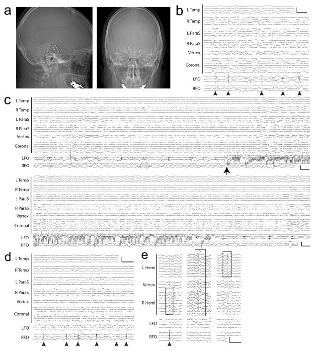
Subclinical mTL seizures and spikes captured with FO electrodes in two AD patients. (A–C): Patient #1. A. Skull x-rays demonstrating radiopaque FO electrodes, with lateral (left) and anterior-posterior (right) views. B. Left mTL spikes (arrowheads), without scalp EEG correlate. Calibration scale: 200uV, 1 sec. C. Electrographic seizure from the left mTL (arrow), without scalp EEG ictal correlate. Panels show continuous EEG spanning 60 seconds. Calibration scale: 150uV, 1 sec. (D–E): Patient #2. D. Right mTL spikes (arrowheads), without scalp EEG correlate. Calibration scale: 200uV, 1 sec. E. Three types of epileptiform discharges. Left: Right mTL spike (arrowhead) with scalp EEG correlate resembling benign epileptiform transient of sleep (box). Middle: Bifrontal spike-wave on scalp EEG (box) without FO correlate. Right: Left temporal sharp wave on scalp EEG (box) without FO correlate. Calibration scale: 200uV, 1 sec. (B–D): Anterior-posterior bipolar montage. L Temp = left temporal (Fp1-F7, F7-T3, T3-T5, T5-O1), R Temp = right temporal (Fp2-F8, F8-T4, T4-T6, T6-O2), L ParaS = left parasagittal (Fp1-F3, F3-C3, C3-P3, P3-O1), R ParaS = right parasagittal (Fp2-F4, F4-C4, C4-P4, P4-O2), Vertex (Fz-Cz, Cz-Pz), Coronal = coronal ring (T1-T3, T3-C3, C3-Cz, Cz-C4, C4-T4, T4-T2, T2-T1), LFO = left FO (LFO1–2, LFO2–3, LFO3–4), RFO = right FO (RFO1–2, RFO2–3, RFO3–4). (E): Referential montage (C2 reference). L Hemi = left hemisphere (Fp1, F7, T1, T3, T5, F3, C3, P3, O1), Vertex (Fz, Cz, Pz), R Hemi = right hemisphere (Fp2, F8, T2, T4, T6, F4, C4, P4, O2), LFO = left FO (LFO1, LFO2, LFO3, LFO4), RFO = right FO (RFO1, RFO2, RFO3, RFO4). Contact 1 is deepest on FOs.
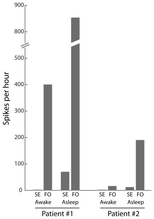
mTL spikes detected on FO electrodes are absent from scalp EEG recordings. Quantification and comparison of spike frequencies simultaneously observed on scalp EEG (SE) and FO electrodes, during wakefulness and sleep, for Patient #1 and #2.
Comment in
-
Let's Talk About Sex: Integrating Sex as a Biological Variable Into Epilepsy Research.Epilepsy Curr. 2018 Sep-Oct;18(5):292-294. doi: 10.5698/1535-7597.18.5.292. Epilepsy Curr. 2018. PMID: 30464725 Free PMC article.
-
Epilepsy and Alzheimer's Disease: Ubiquitous Entities Subject to the Same Cosmic Forces but on Different Astral Planes.Epilepsy Curr. 2018 Sep-Oct;18(5):295-297. doi: 10.5698/1535-7597.18.5.295. Epilepsy Curr. 2018. PMID: 30464726 Free PMC article. No abstract available.
References
-
- Alzheimer A. Über einen eigenartigen schweren Erkrankungsprozeβ der Hirnrinde. Neurol Cent. 1906;23:1129–36.
-
- Blocq P, Marinesco G. Sur les lesions et la pathogenie de l’epilepsie dite essentielle. La Sem Medicale. 1892;12:445–446.
Publication types
MeSH terms
Grants and funding
LinkOut - more resources
Full Text Sources
Other Literature Sources
Medical
