. 2017 Jun;35(6):569-576.
doi: 10.1038/nbt.3836. Epub 2017 May 1.

In vivo genome editing and organoid transplantation models of colorectal cancer and metastasis

Affiliations

In vivo genome editing and organoid transplantation models of colorectal cancer and metastasis

Jatin Roper et al. Nat Biotechnol. 2017 Jun.

Erratum in

  • Corrigendum: In vivo genome editing and organoid transplantation models of colorectal cancer and metastasis.
    Roper J, Tammela T, Cetinbas NM, Akkad A, Roghanian A, Rickelt S, Almeqdadi M, Wu K, Oberli MA, Sánchez-Rivera F, Park YK, Liang X, Eng G, Taylor MS, Azimi R, Kedrin D, Neupane R, Beyaz S, Sicinska ET, Suarez Y, Yoo J, Chen L, Zukerberg L, Katajisto P, Deshpande V, Bass AJ, Tsichlis PN, Lees J, Langer R, Hynes RO, Chen J, Bhutkar A, Jacks T, Yilmaz ÖH. Roper J, et al. Nat Biotechnol. 2017 Dec 8;35(12):1211. doi: 10.1038/nbt1217-1211a. Nat Biotechnol. 2017. PMID: 29220013

Abstract

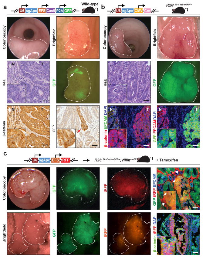
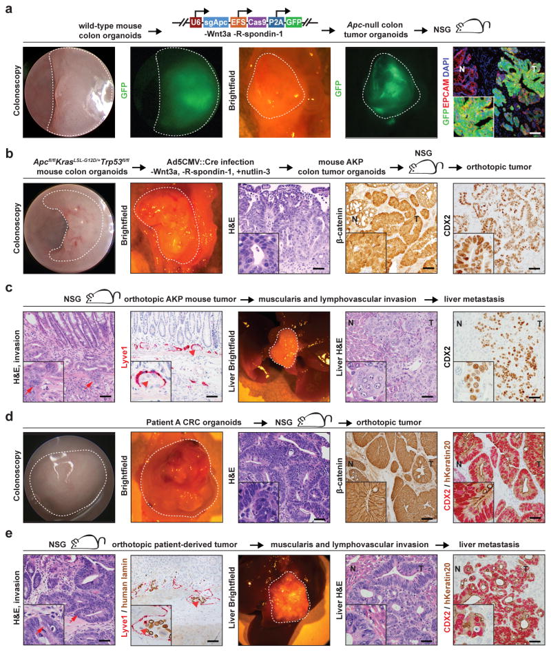
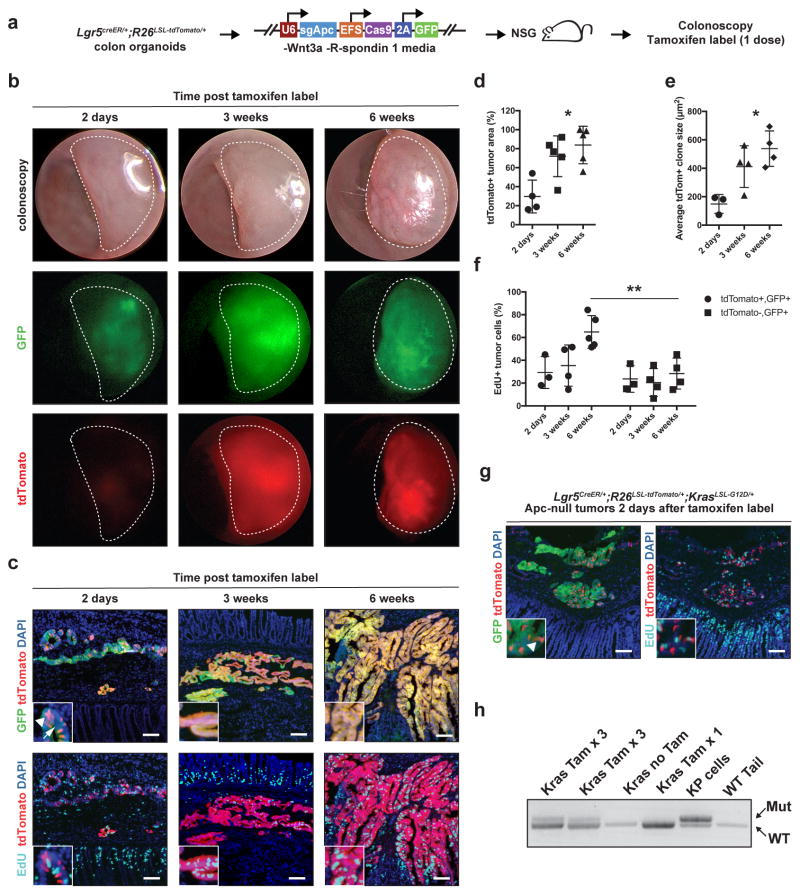
In vivo interrogation of the function of genes implicated in tumorigenesis is limited by the need to generate and cross germline mutant mice. Here we describe approaches to model colorectal cancer (CRC) and metastasis, which rely on in situ gene editing and orthotopic organoid transplantation in mice without cancer-predisposing mutations. Autochthonous tumor formation is induced by CRISPR-Cas9-based editing of the Apc and Trp53 tumor suppressor genes in colon epithelial cells and by orthotopic transplantation of Apc-edited colon organoids. ApcΔ/Δ;KrasG12D/+;Trp53Δ/Δ (AKP) mouse colon organoids and human CRC organoids engraft in the distal colon and metastasize to the liver. Finally, we apply the orthotopic transplantation model to characterize the clonal dynamics of Lgr5+ stem cells and demonstrate sequential activation of an oncogene in established colon adenomas. These experimental systems enable rapid in vivo characterization of cancer-associated genes and reproduce the entire spectrum of tumor progression and metastasis.


Conflict of interest statement

Competing Financial Interests Statement: The authors have no competing financial interests

Figures

Figure 1
Figure 1. CRISPR-Cas9-based in situ Apc editing in the colon epithelium induces adenoma formation
(a) Tumors in wild-type mice following mucosal injection with 1,000 transforming units (TU) per μl of U6::sgApc-EFS::Cas9-P2A-GFP lentivirus. Tumors are indicated with white light colonoscopy, brightfield necropsy, GFP fluorescence necropsy, Hematoxylin and eosin (H&E) staining, and β-catenin immunohistochemistry. Note patchy GFP expression in tumors (arrow). (b) Tumors in Rosa26LSL-Cas9-eGFP/+ mice after mucosal delivery of a lentiviruses encoding an sgRNA against Apc and Cre recombinase (U6::sgApc-CMV::Cre, 10,000 TU/μl) (white light colonoscopy, brightfield necropsy, and GFP fluorescence necropsy; H&E immunohistochemistry and GFP/β-catenin immunofluorescence). GFP tumor fluorescence indicates Cre-induced expression of Cas9 and eGFP from the Rosa26 locus. (c) Tumorigenesis in Rosa26LSL-Cas9-eGFP/+;VillinCreER mice treated with tamoxifen and then injected with lentiviruses encoding sgApc and turboRFP (U6::sgApc-EFS::turboRFP, 10,000 TU/μl) into the colon mucosa (white light/GFP/turboRFP fluorescence colonoscopy and brightfield/GFP/turboRFP fluorescence necropsy; GFP/turboRFP immunofluorescence). Arrowheads indicate turboRFP expression in stromal cells. Arrows point to GFP/turboRFP dual-positive tumor cells. Histology images are 20X and insets are 60X (Scale bar: 200 μm). Dotted lines indicate tumors. (tRFP: turboRFP; R26: Rosa26; N: normal; T: tumor).
Figure 2
Figure 2. Orthotopic transplantation models of mouse and patient-derived colorectal cancer
(a) Tumors in the distal colons of NSG mice following orthotopic transplantation of wild-type colon organoids infected with U6::sgApc-EFS::Cas9-P2A-GFP lentivirus. Tumors are visualized with white light/GFP fluorescence colonoscopy, white light/GFP fluorescence necropsy, and GFP immunofluorescence. (b) Tumors induced in NSG mice by orthotopic transplantation of ApcΔ/ Δ, KrasG12D/+, Trp53Δ/ Δ (AKP) colon organoids. Tumors are imaged with colonoscopy, necropsy, hematoxylin and eosin (H&E) staining, β-catenin immunohistochemistry, and CDX2 immunohistochemistry. (c) Local invasion and liver metastases of engrafted AKP colon organoid tumors, as indicated H&E and LYVE1 immunohistochemistry of primary colon tumors, necropsy, and CDX2 immunohistochemistry of liver metastases. Arrows indicate invasion of the muscularis propria and arrowheads demonstrate tumor invasion of a LYVE1-positive lymphatic vessel. (d) Tumors in NSG mice following orthotopic transplantation of patient-derived CRC organoids. Tumors are demonstrated with colonoscopy, necropsy, H&E staining, β-catenin immunohistochemistry, and CDX2/human Keratin20 immunohistochemistry. (e) Local invasion and liver metastases of patient-derived organoid orthotopic tumors are demonstrated by H&E staining, LYVE1 immunohistochemistry, liver necropsy, and CDX2/human Keratin20 immunohistochemistry. Arrows denote invasion of the muscularis and arrowheads indicate tumor invasion of a LYVE1 positive vessel. Histology images are 20X and insets are 60X (Scale bar: 200 μm). Dotted lines indicate tumors. (NSG: nod SCID gamma; T: tumor; N: normal colon; hKeratin20: human Keratin 20).
Figure 3
Figure 3. Lgr5 cell lineage tracing and sequential mutagenesis in established orthotopic colon adenomas
(a) Colon organoids derived from Lgr5CreER/+;Rosa26LSL-tdTomato/+ mice were subjected to CRISPR-Cas9-based Apc editing with U6::sgApc-EFS::Cas9-P2A-GFP infection, and then orthotopically transplanted into NSG mice to generate Apc-null in vivo tumors. Tumor-bearing mice received one dose of tamoxifen to label Lgr5+ cells and their progeny with tdTomato and were evaluated 2 days, 3 weeks, or 6 weeks later. Proliferating cells were marked with a 5-ethynyl-2′-deoxyuridine (EdU) pulse 4 hours before sacrifice. (b) In vivo tumor imaging by white light, GFP fluorescence, and tdTomato fluorescence colonoscopy (dotted lines) at 2 days, 3 and 6 weeks after cell labeling. (c) GFP, tdTomato, and EdU immunofluorescence images of orthotopic tumors. Arrowhead indicates cell labeling with tdTomato at 2 days post label (white arrowhead). Arrow points to GFP-negative tumor areas that suggest mosaic lentiviral silencing. Immunofluorescence images were analyzed for: (d) tdTomato+ tumor area relative to total GFP+ tumor area; (e) average tdTomato+ clone size (i.e., total tdTomato+ area / total clone number); and (f) proportion of EdU+ proliferating tdTomato+, GFP+ tumor cells vs. EdU+ proliferating tdTomato−, GFP+ tumor cells at 6 weeks post labelling. (N=3 or 4 tumors for each group and time point). (g) Clonal activation of tdTomato+ (arrowhead) 2 days after tamoxifen administration to mice bearing orthotopic Apc-null Lgr5CreER/+;Rosa26LSL-tdTomato/+;KrasLSL-G12D/+ tumors; (h) Analysis of recombination of the lox-stop-lox (LSL) cassette at the mutant KrasLSL-G12D/+ locus in response to tamoxifen-mediated activation of CreER. Tam x 3 indicates tumors where mice received 3 pulses of tamoxifen, and Tam x1 where one pulse was given. Tissues were harvested 48 hours after the last pulse of tamoxifen. Wild-type mouse tail DNA was used as a negative control, and KrasG12D/+;Trp53Δ/ Δ lung adenocarcinoma cell line DNA (KP cells) as a positive control. *P<0.005 (One-way ANOVA), **P=0.01 (Student’s T-test). (R26: Rosa26; NSG: nod SCID gamma; tdTom: tdTomato)

Comment in

Similar articles

Cited by

References

    1. Cancer Genome Atlas Network. Comprehensive molecular characterization of human colon and rectal cancer. Nature. 2012;487:330–337. - PMC - PubMed
    1. Seshagiri S, et al. Recurrent R-spondin fusions in colon cancer. Nature. 2012;488:660–664. - PMC - PubMed
    1. Moser AR, Pitot HC, Dove WF. A dominant mutation that predisposes to multiple intestinal neoplasia in the mouse. Science. 1990;247:322–324. - PubMed
    1. Su LK, et al. Multiple intestinal neoplasia caused by a mutation in the murine homolog of the APC gene. Science. 1992;256:668–670. - PubMed
    1. Roper J, Hung KE. Priceless GEMMs: genetically engineered mouse models for colorectal cancer drug development. Trends Pharmacol Sci. 2012;33:449–455. - PubMed

Publication types