. 2017 Jun;35(6):577-582.
doi: 10.1038/nbt.3837. Epub 2017 May 1.

Transplantation of engineered organoids enables rapid generation of metastatic mouse models of colorectal cancer

Affiliations

Transplantation of engineered organoids enables rapid generation of metastatic mouse models of colorectal cancer

Kevin P O'Rourke et al. Nat Biotechnol. 2017 Jun.

Abstract

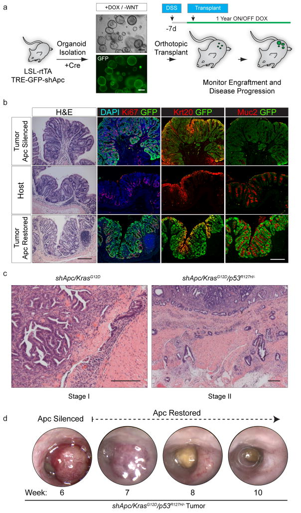
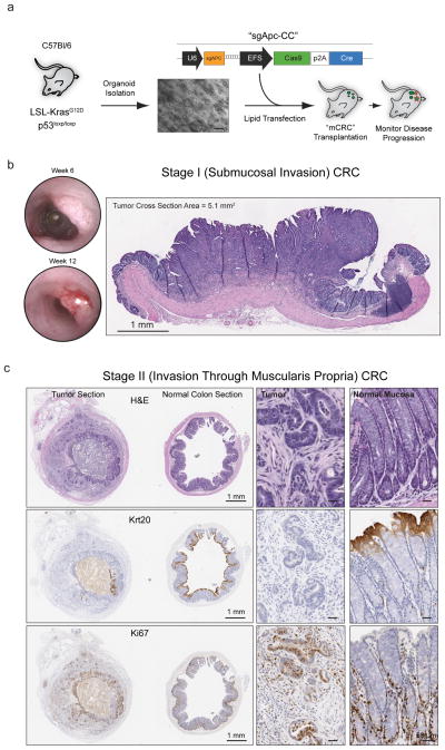
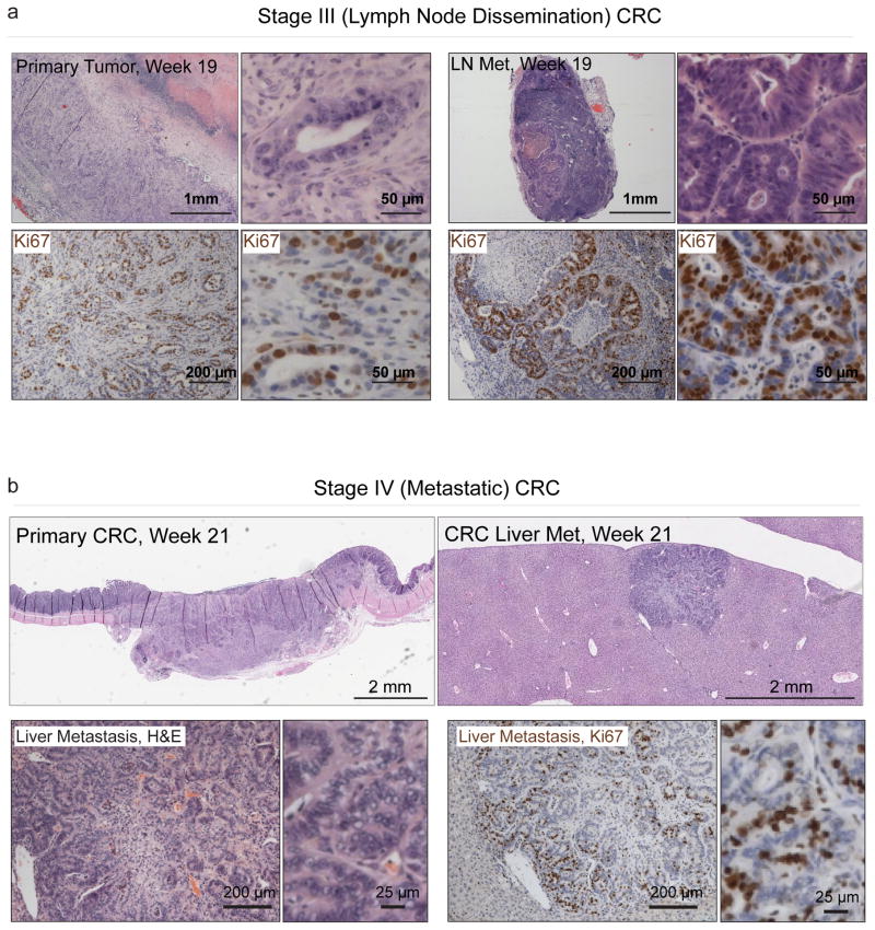
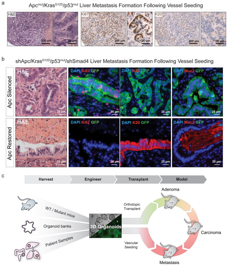
Colorectal cancer (CRC) is a leading cause of death in the developed world, yet facile preclinical models that mimic the natural stages of CRC progression are lacking. Through the orthotopic engraftment of colon organoids we describe a broadly usable immunocompetent CRC model that recapitulates the entire adenoma-adenocarcinoma-metastasis axis in vivo. The engraftment procedure takes less than 5 minutes, shows efficient tumor engraftment in two-thirds of mice, and can be achieved using organoids derived from genetically engineered mouse models (GEMMs), wild-type organoids engineered ex vivo, or from patient-derived human CRC organoids. In this model, we describe the genotype and time-dependent progression of CRCs from adenocarcinoma (6 weeks), to local disseminated disease (11-12 weeks), and spontaneous metastasis (>20 weeks). Further, we use the system to show that loss of dysregulated Wnt signaling is critical for the progression of disseminated CRCs. Thus, our approach provides a fast and flexible means to produce tailored CRC mouse models for genetic studies and pre-clinical investigation.


Conflict of interest statement

Competing Financial Interests: The authors declare no competing financial interests.

Figures

Figure 1
Figure 1
Transplanted organoids can recapitulate autochthonous GEMMs a: Schematic depiction of the protocol used to generate organoid engraftments to model Apc loss in the colon. b. Histochemical (H&E) and immunofluorescent (Ki67, Krt20, Muc2, GFP) stains of an engrafted shApc adenoma (top row) and neighboring normal host mucosa (middle row), and long-term (29 weeks) Apc-restored (bottom row). c. H&E stain of an shApc/KrasG12D tumor 8 weeks post transplant (left), H&E stain of an shApc/KrasG12D/p53R172H/− tumor 16.5 weeks post transplant (right). d. Serial endoscopies of an shApc/KrasG12D/p53R172H/− tumor that was maintained ON Dox (Apc Silenced) for 6 weeks, and then longitudinally followed OFF Dox (Apc restored). Scale bars are 200 μm.
Figure 2
Figure 2
Transplanted organoids create focal colorectal tumors. a. Schematic depiction of the protocol used to generate Apcmut/KrasG12D/p53mut organoids for syngeneic orthotopic engraftment. b. Serial endoscopies of an Apcmut/KrasG12D/p53mut transplant growing as a polypoid mass in the mucosal layer of an engrafted host (left), and H&E stained cross-section of an Apcmut/KrasG12D/p53mut tumor (7 weeks post transplant). c. Histochemical (H&E) and immunohistochemical (Krt20, Ki67) stained sections of tumor (left) and normal (right) tissue of a Stage II CRC tumor collected 75 days post transplant. Whole slide scan images are presented on the left (Scale Bar, 1mm), with higher resolution insets on the right (Scale Bar, 50 μm).
Figure 3
Figure 3
Orthotopically transplanted organoids progress to metastatic CRC. a. Histochemical (H&E, top) and immunohistochemical (Ki67, bottom) of a T3 primary tumor, 137 days post transplant, and a caudal lymph node metastasis (right). b. H&E stained sections (top) of a primary T3 CRC tumor 21 weeks post transplant, and a liver metastasis harvested from the same animal (low res slide scan, top right, higher magnification, bottom left), and immunohistochemical (Ki67) stained section of the liver met (bottom right).
Figure 4
Figure 4
Dysregulated Wnt signaling is required to sustain advanced CRC.a. Histochemical (H&E, left) and immunohistochemical (Ki67, middle, Krt20, right) stains of sections from an Apcmut/KrasG12D/p53mut liver metastasis that formed 10 weeks after intra-splenic injection. b. Histochemical (H&E) and immunofluorescent (GFP, Ki67, Krt20, Muc2, Dapi) stains of liver tumors in animals maintained on dox for 10 weeks (“Apc silenced”),” see also Supplementary Fig. 19f), and of liver tumors maintained on dox for 10 weeks, then off dox for 3 weeks (“Apc Restoration,” bottom). c. A schematic depicting the two-tiered approach for ex vivo manipulation of organoids, sourced from murine models, tissue banks or human samples, engineered to further alter genotype, and transplanted into the colon mucosa by DSS treatment/pipette enema. In parallel organoids can be seeded in the vasculature to model metastatic CRC growth.

Comment in

References

    1. Fearon ER, Vogelstein B. A genetic model for colorectal tumorigenesis. Cell. 1990;61:759–767. - PubMed
    1. Markowitz SD, Bertagnolli MM. Molecular origins of cancer: Molecular basis of colorectal cancer. New England Journal of Medicine. 2009;361:2449–2460. - PMC - PubMed
    1. Taketo MM, Edelmann W. Mouse Models of Colon Cancer. YGAST. 2009;136:780–798. - PubMed
    1. Heijstek MW, Kranenburg O, Borel Rinkes IHM. Mouse Models of Colorectal Cancer and Liver Metastases. Digestive Surgery. 2005;22:16–25. - PubMed
    1. Oh BY, Hong HK, Lee WY, Cho YB. Animal models of colorectal cancer with liver metastasis. Cancer Lett. 2016 - PubMed

Publication types