. 2017 Jun;35(6):583-589.
doi: 10.1038/nbt.3840. Epub 2017 May 1.

Generation of inner ear organoids containing functional hair cells from human pluripotent stem cells

Affiliations

Generation of inner ear organoids containing functional hair cells from human pluripotent stem cells

Karl R Koehler et al. Nat Biotechnol. 2017 Jun.

Abstract

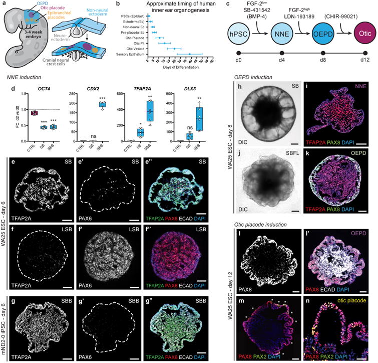
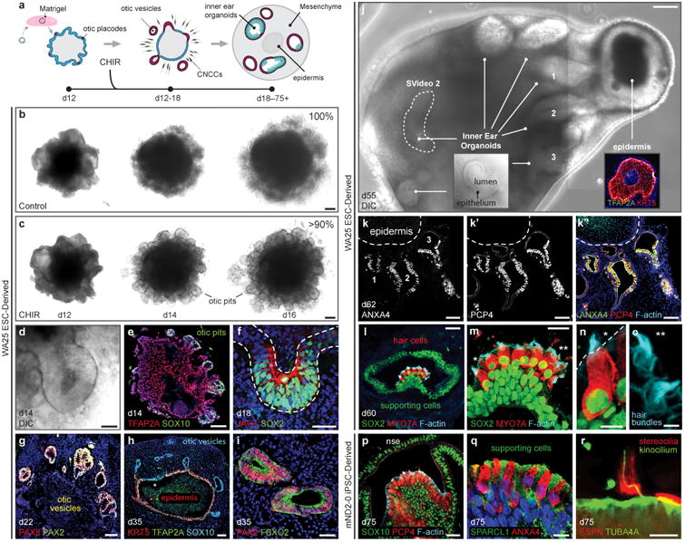
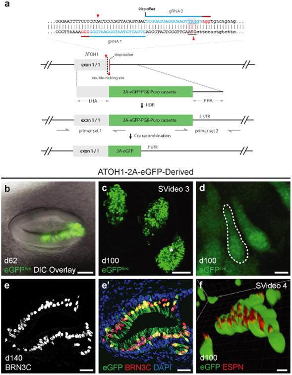
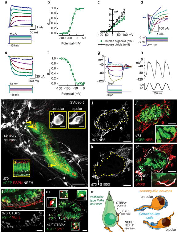
The derivation of human inner ear tissue from pluripotent stem cells would enable in vitro screening of drug candidates for the treatment of hearing and balance dysfunction and may provide a source of cells for cell-based therapies of the inner ear. Here we report a method for differentiating human pluripotent stem cells to inner ear organoids that harbor functional hair cells. Using a three-dimensional culture system, we modulate TGF, BMP, FGF, and WNT signaling to generate multiple otic-vesicle-like structures from a single stem-cell aggregate. Over 2 months, the vesicles develop into inner ear organoids with sensory epithelia that are innervated by sensory neurons. Additionally, using CRISPR-Cas9, we generate an ATOH1-2A-eGFP cell line to detect hair cell induction and demonstrate that derived hair cells exhibit electrophysiological properties similar to those of native sensory hair cells. Our culture system should facilitate the study of human inner ear development and research on therapies for diseases of the inner ear.


Figures

Figure 1
Figure 1
Step-wise induction of otic placode-like epithelia. a, Overview of mammalian ectoderm development in the otic placode cranial region. b, Timeline for key events of human otic induction. Day 0 on the timeline indicates the approximate stage of development represented by hPSC: ∼12 dpc. c, Differentiation strategy for non-neural ectoderm (NNE), otic-epibranchial progenitor domain (OEPD), and otic placode induction. Potentially optional or cell line-dependent treatments are denoted in parentheses. d, qPCR analysis on day 2 of differentiation of WA25 cell aggregates treated with DMSO (Control), 10 μM SB, or 10 μM SB + 10 ng/ml BMP4, denoted as SBB. Gene expression was normalized to undifferentiated hESCs; n = 3 biological samples, 2 technical repeats; *P<0.05, **P<0.01, ***P<0.001; error bars = max/min. e, f, Representative TFAP2A, ECAD, and PAX6 expression in WA25 aggregate treated with 10 μM SB or with 200 nM LDN + 10 μM SB for 6 days. g, TFAP2A, ECAD, and PAX6 expression in mND2-0 iPSCs treated with 10 μM SB + 2.5 ng/ml BMP4 (SBB) on day 6. h, i, Representative image of a SB-treated WA25 aggregate on day 8: live (h) and immunostained with PAX8 and TFAP2A antibodies (i). When comparing morphology in panels (h) and (i) note that the outer-epithelium crumples into the aggregate core during the cryosectioning process. j, k, Representative image of a SB-treated WA25 aggregate on day 8 after treatment with 50 ng/ml FGF-2 and 200 nM LDN (SBFL) on day 4: live (j) and immunostained with PAX8 and TFAP2A antibodies (k). l-n, WA25 SBFL-treated aggregates on day 12. The outer-epithelium contains PAX8+ ECAD+ cells (l) and occasional patches of PAX8+ PAX2+ otic placode-like cells (m, n). The specimens shown were treated with 25 μl of additional CDM on day 8. Scale bars, 100 μm (e-m), 50 μm (n).
Figure 2
Figure 2
Wnt signaling activation initiates self-organization and maturation of inner ear organoids containing vestibular-like hair cells. a, Inner ear organoid induction strategy. Day 12 aggregates were embedded in Matrigel droplets to support vesicle formation. b-d, In CHIR-treated samples, but not DMSO (Control) samples, otic pit-like structures evaginate from the outer-epithelium (d). e-i, Between days 14-35, pits and vesicles expressed otic specific markers, such as SOX10, SOX2, JAG1, PAX8, PAX2, and FBXO2. The epithelium from which vesicles arise begins to express the epidermal keratinocyte marker KRT5 by day 35 (h). j, By day 40-60, the aggregates contain multiple organoids and, typically, a single epidermal unit visible under DIC imaging. Inner ear organoids are distinguishable by a defined epithelium with ∼25-40 μm apparent thickness and a lumen (j inset). k, Inner ear organoids are typically oriented around the epidermal unit and contain sensory epithelia with ANXA4+ PCP4+ hair cells. The luminal surface of organoids is actin-rich, as denoted by phalloidin staining (k″). l-o, Hair cells are MYO7A+ SOX2+, and supporting cells are SOX2+. F-actin-rich hair bundles protrude from the hair cells into the lumen (n, o; asterisks denote hair bundle location in m). p, q, mND2-0 iPSC-derived sensory epithelia have a similar morphology to WA25 hESC-derived sensory epithelia and contain PCP4+ ANXA4+ hair cells. SOX10 is expressed throughout the supporting and non-sensory epithelial cell populations, but not in hair cells (p). Supporting cells express the utricle supporting cell marker SPARCL1 (q). r, Hair cells in organoids have ESPN+ hair bundles with a single acetylated-tubulin (TUBA4A)+ kinocilium. Scale bars, 200 μm (j), 100 μm (b, c, e), 50 μm (d, g, h, k, l), 25 μm (f, i, m, p), 10 μm (n, q), 5 μm (r), 2.5 μm (o).
Figure 3
Figure 3
Development of an ATOH1 fluorescent reporter hESC line for tracking hair cell induction. a, ATOH1-2A-eGFP CRISPR design. The two guide RNAs (blue, with PAM sequence in red) direct Cas9n to make two nicks (red triangles) near the stop codon (underlined with pink background) of ATOH1. The resulting DNA double strand break is repaired by the donor vector, which has a 2A-eGFP-PGK-Puro cassette and 1kb left and right homology arms (LHA and RHA). The LoxP-flanked PGK-Puro sub-cassette is subsequently removed by Cre recombinase. In ATOH1 expressing hair cells, eGFP is transcribed along with ATOH1. b-d, Representative live cell images of eGFP+ hair cells in 62- and 100-day-old inner ear organoids. The asterisk in panel (c) denotes the approximate location of the hair cells in panel (d). e, Expression of BRN3C in 140-day-old eGFP+ hair cells. f, Expression of ESPN in the hair bundles of 100-day-old eGFP+ hair cells. Scale bars, 100 μm (c), 50 μm (b), 25 μm (e), 5 μm (d, f).
Figure 4
Figure 4
hESC-derived hair cells have similar electrophysiological properties as native hair cells and form synapse-like contacts with sensory neurons. a, Family of outward rectifier potassium currents recorded from a human organoid hair cell (d64), evoked by the series of voltage steps shown below. b, The outward currents had an activation range that was well-fitted by a Boltzmann equation (line) with voltage of half maximal activation of −31 mV. c, Mean (± S.E.) maximal current-voltage relationships for seven human organoid hair cells (d64-d67) and eight mouse utricle type II hair cells. For current-voltage relations, we averaged data from hair cells with large currents over 2 nA. d, Family of rapidly-activating, rapidly-inactivating, inward currents evoked by the depolarizing steps shown below. e, Family of slowly-activating, non-inactivating inward currents (d64) evoked by hyperpolarizing steps, shown below. f, Activation curve for the current family shown in panel e, fitted by a Boltzmann equation (line) with a voltage of half maximal activation of −71 mV. g, Family of membrane responses (d64) recorded in current-clamp mode, evoked by the current injection protocol shown below. h, Membrane response (d65) to three cycles of a 5-Hz sine wave stimulus (below). i, 3D projection of eGFP+ hair cells with ESPN+ hair bundles surrounded by clusters of sensory-like neurons. Insets 1 and 2 demonstrate the two neuron morphologies observed: unipolar and bipolar. Inset 3 demonstrates hair cell morphology and NEFH+ neurites in the sensory epithelium. j, Representative image of NEFL+ neurons innervating an organoid sensory epithelium. k, S100β+ Schwann-like cells associate with neuronal soma and appear to myelinate NEFL+ neuronal processes. l, NEFL+ neuronal processes infiltrate the epithelium and are closely associated with CTBP2+ puncta at the base of eGFP+ hair cells. m, CTBP2+ puncta are co-localized with SYP+ puncta. n, Summary of neurogenesis analysis. Scale bars, 100 μm (i), 25 μm (j, k), 10 μm (l, m), 5 μm (i insets).

Comment in

References

    1. Géléoc GSG, Holt JR. Sound strategies for hearing restoration. Science. 2014;344:1241062. - PMC - PubMed
    1. Rosenhall U. Degenerative patterns in the aging human vestibular neuro-epithelia. Acta oto-laryngologica. 1973;76:208–220. - PubMed
    1. Müller U, Barr-Gillespie PG. New treatment options for hearing loss. Nat Rev Drug Discov. 2015;14:346–365. - PubMed
    1. Sergeyenko Y, Lall K, Liberman MC, Kujawa SG. Age-Related Cochlear Synaptopathy: An Early-Onset Contributor to Auditory Functional Decline. 2013;33:13686–13694. - PMC - PubMed
    1. Koehler KR, Hashino E. 3D mouse embryonic stem cell culture for generating inner ear organoids. Nature Protocols. 2014;9:1229–1244. - PMC - PubMed

Publication types

MeSH terms